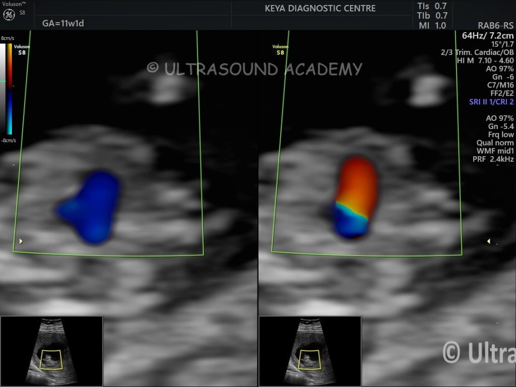
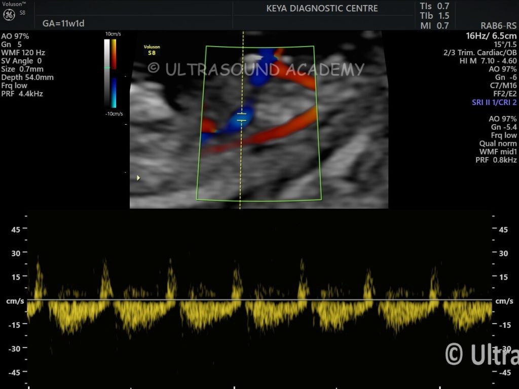
Findings and Description- Normal size and flow in both ventricles. LVOT showing antegrade flow . RVOT appears dilated and showing reversal of flow direction S/O Bidirectional flow. Bidirectional flow on Spectral Doppler also noted.
4CH view may be normal in early scans
Outflow tracts can also of normal caliber in early scans
Typical bidirectional flow is obvious finding on Color Doppler and spectral Doppler
During later stages of gestation dilatation of main pulmonary artery and its branches becomes more
evident.
Differential Diagnosis
-Tetralogy of Fallot
-Primary Pulmonary valve dysplasis/stenosis
-Pulmonary atresia with VSD
Key Features:
Fetal Absent Pulmonary Valve Syndrome (APVS) is a rare and serious congenital heart defect characterized by
– Complete absence or severe underdevelopment of pulmonary valve
– Significant dilation of pulmonary arteries on 2nd trimester scans onwards.
– Often associated with other cardiac malformations
– High mortality rate, especially in severe cases
Pathophysiology:
– Disrupts normal blood flow from right ventricle to lungs
– Causes massive pulmonary artery dilation
– Leads to potential respiratory distress and cardiac compromise
Diagnostic Characteristics:
– Can be detected on cardiac scanning as early as 11- 12 wks.
– Marked pulmonary artery and branch arteries are significantly enlargement on 2nd trimester scan
– Potential right ventricular dysfunction
– Possible bronchial compression due to arterial dilation
Potential Complications:
– Respiratory insufficiency
– Cardiac failure
– Potential need for early surgical intervention
– Risk of significant morbidity and mortality
Management:
– Requires multidisciplinary approach
– Potential surgical repair or valve replacement
– Close cardiac and respiratory monitoring
– Individualized treatment based on specific anatomical findings
Prognosis varies significantly depending on severity and associated cardiac anomalies, with some cases requiring immediate intervention after birth.