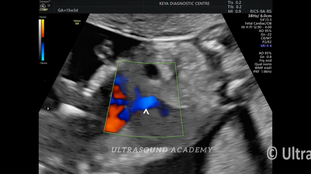
NORMAL DUCTUS VENOSUS AT 13 WKS
A 24 weeks primigravida, came for NT scan at 13 weeks of gestation.
Findings:
Normal ductus venosus shown on colour doppler imaging showing aliasing.
Discussion:
In first-trimester done as a screening for aneuploidic anomalies
In second and third-trimester as a screening for IUGR or for fetal cardiac compromise.
Reversed a-wave in ductus venosus between 16-20 weeks’ gestation is associated with increased risk of trisomy 21, preeclampsia, and GDM.
4