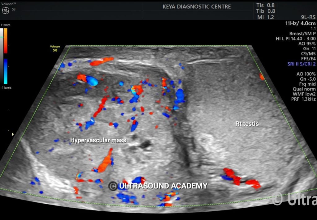
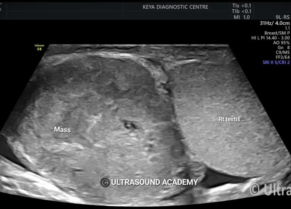
40 year male with right scrotal swelling for 6 months, complaining of pain since last few days ,
Referred to us for USG Scrotum.
Findings & Discussion:
There is large , well defined , predominantly solid , isoechoic mass lesion is noted in the right para-testicular region , arising from the head of epididymis.
Few tiny cystic spaces seen at the center of the lesion.
No evidence of calcification seen within the lesion.
On color Doppler , the lesion shows significantly increased vascularity .
Right testis appears unremarkable and seen separately from the above mentioned lesion.
PET CT showed significantly high uptake in the lesion.
Histopathology was suggestive of High grade Undifferentiated malignancy
Ultrasound is a key imaging modality for evaluating epididymal malignancy and differentiating it from benign conditions.
Ultrasound Features of Epididymal Malignancy
Hypoechoic Mass
Malignant tumors usually appear as solid, hypoechoic masses within the epididymis.
May have irregular borders.
Increased Vascularity on Doppler
Malignant lesions often show increased blood flow on color Doppler.
Benign lesions, like epididymal cysts or spermatoceles, typically lack internal vascularity.
Heterogeneous Echotexture
May contain cystic and solid components.
Can have areas of necrosis or hemorrhage.
Size and Growth Pattern
Rapidly enlarging epididymal masses are more concerning for malignancy.
Local invasion into adjacent testicular or scrotal structures suggests an aggressive tumor.
Calcifications
Some epididymal malignancies may show microcalcifications.
Associated Findings
Possible reactive hydrocele.
Involvement of spermatic cord or testis in advanced cases.
Common Malignant Tumors of the Epididymis
Adenomatoid Tumor (usually benign but can be aggressive)
Leiomyosarcoma (rare but aggressive)
Metastases (secondary tumors, often from the prostate, lung, or kidney)
Differential Diagnoses (Benign Conditions)
Epididymal cysts (anechoic, well-defined, no vascularity)
Spermatocele (cystic with internal echoes)
Epididymitis (enlarged, hypervascular, painful)
Tuberculosis of the epididymis (heterogeneous, nodular)
Next Steps if Malignancy is Suspected
Scrotal MRI (for better tissue characterization)
Tumor Markers (AFP, β-hCG, LDH for testicular malignancy involvement)
Biopsy or Surgical Excision (confirmatory diagnosis)