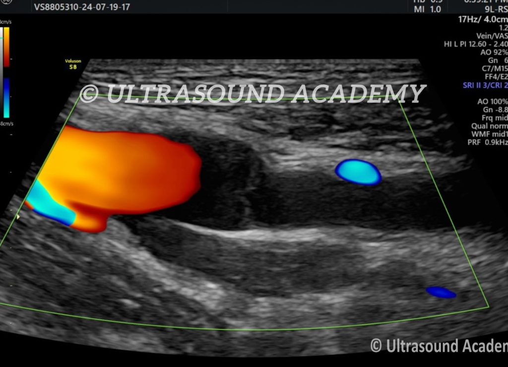
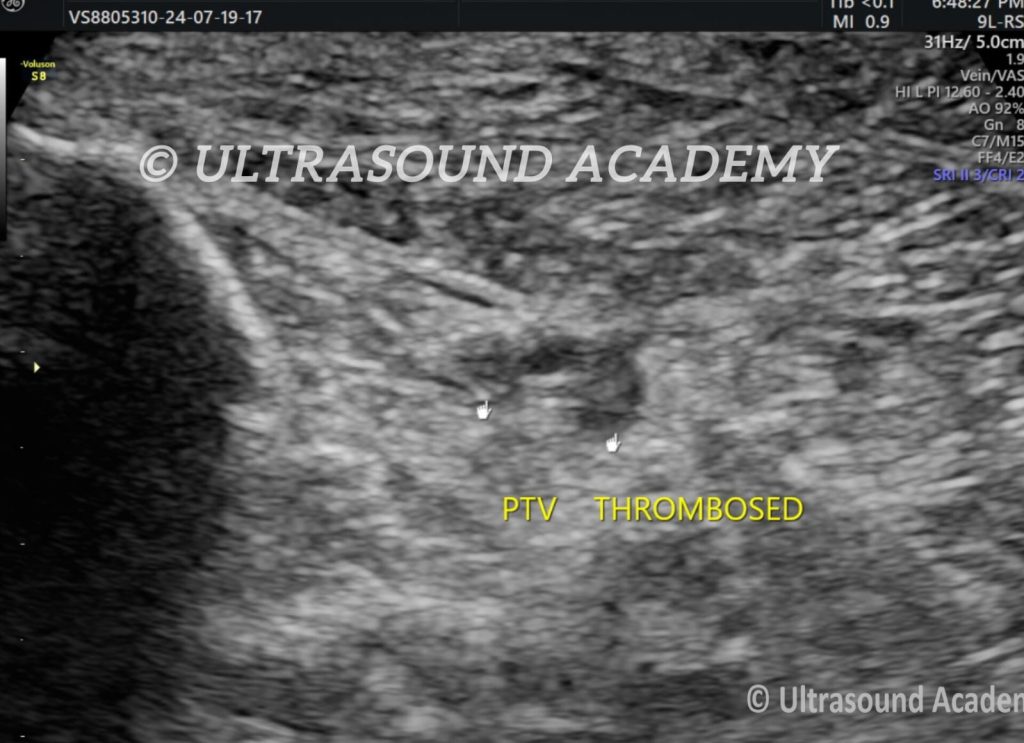
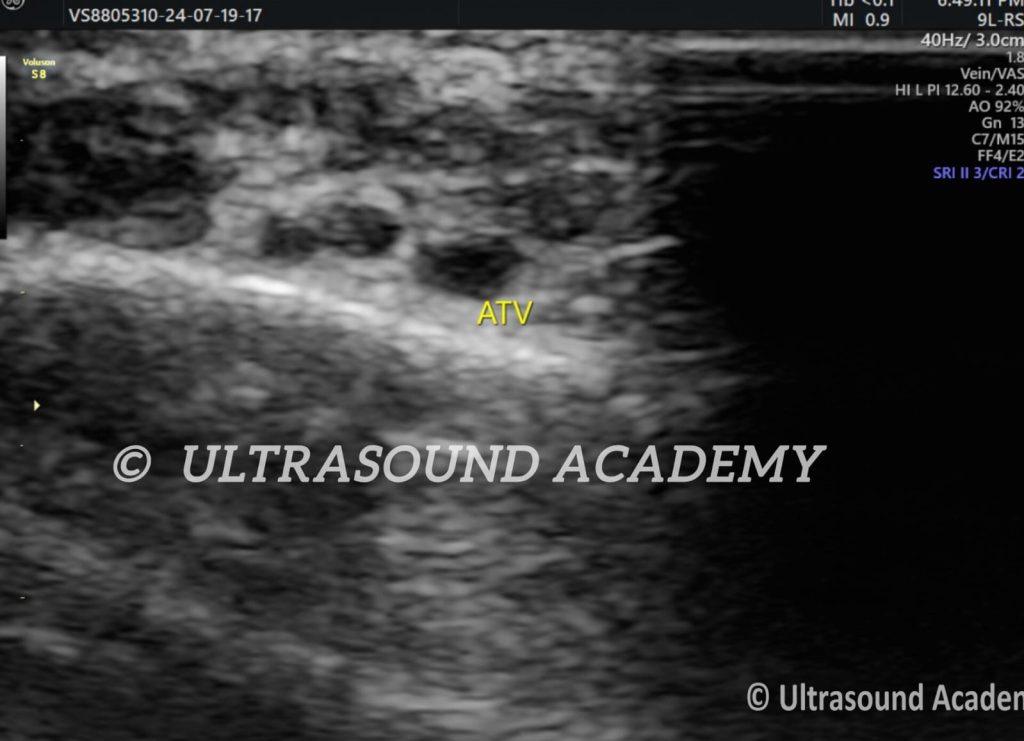
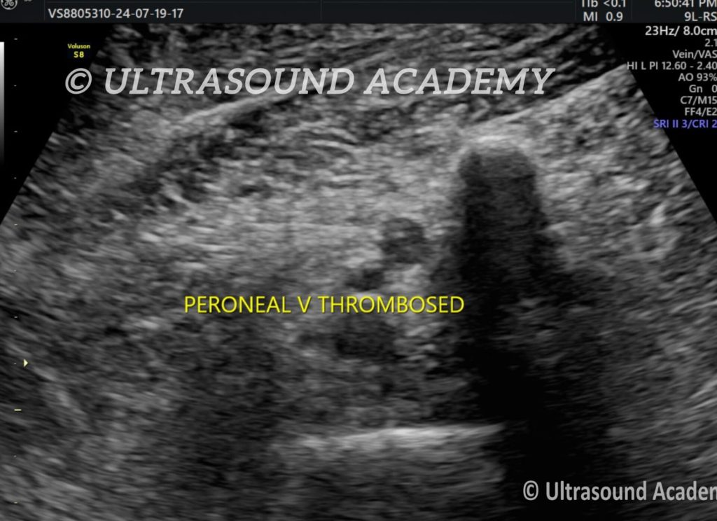
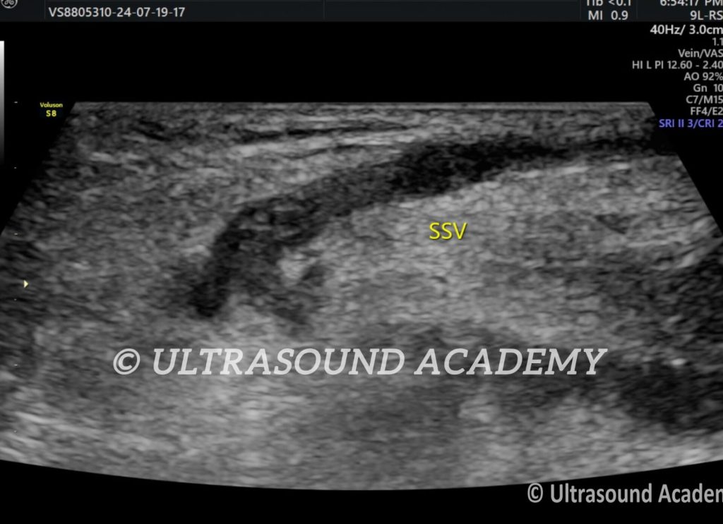
USG FINDINGS: Acute echogenic thrombus is seen in lower limb veins extending from distal part of Superficial Femoral Vein, Popliteal Vein, ATV, PTV, personal veins as well as SSV. Thrombosis Vein appears enlarged in caliber and shows loss of compressibility. Evaluation of Proximal extent of thrombus into abdominal veins is very important for treatment.
A Venous Doppler ultrasound of the lower limb is a non-invasive diagnostic test that uses sound waves to evaluate blood flow in the veins. It is commonly used to assess venous disorders, such as deep vein thrombosis (DVT), venous insufficiency, or varicose veins.
Purpose
Deep Vein Thrombosis (DVT):
Detect blood clots in the deep veins of the leg.
Venous Insufficiency:
Evaluate valve function in the veins and identify backward blood flow (reflux).
Varicose Veins:
Assess the severity and location of abnormal vein dilation.
Post-Thrombotic Syndrome:
Monitor vein damage or complications from prior DVT.
Pre-Surgical Evaluation:
Guide vein mapping for procedures like varicose vein surgery or bypass grafting.
Normal Flow:
Blood should flow smoothly and unidirectionally toward the heart.
Obstruction:
Thrombus or narrowing within the vein.
Reflux:
Reversal of blood flow due to incompetent venous valves.
Vein Compressibility:
Healthy veins compress fully under pressure, while clotted veins do not.
Key Findings
DVT:
Presence of a thrombus (clot), typically shown as a hypoechoic or echogenic mass.
Non-compressibility of the vein.
Venous Insufficiency:
Retrograde blood flow during the Valsalva maneuver or on standing.
Varicose Veins:
Enlarged, tortuous veins with reflux.
Chronic Venous Changes:
Thickened vein walls, decreased vein elasticity, or scarring.
Advantages
Non-invasive and painless.
No radiation exposure.
Real-time dynamic assessment of blood flow.
Limitations
Operator-dependent; accuracy depends on the skill of the sonographer.
May be less sensitive in detecting pelvic vein thrombi or very small clots.
Ultrasound Findings in Venous Doppler of Lower Limb
Normal Findings
Vein Compressibility:
Veins should completely compress when pressure is applied with the transducer.
Indicates no thrombus is present.
Flow Direction:
Blood flows toward the heart in a normal venous system.
Augmentation of Flow:
Increased flow velocity is seen with distal compression of the limb.
Color Doppler:
Uniform color pattern showing laminar flow.
Abnormal Findings
Deep Vein Thrombosis (DVT):
Echogenic Material: Hypoechoic or echogenic thrombus within the vein.
Non-Compressibility: Inability to compress the vein when pressure is applied.
Absence of Flow: No color Doppler signals in the affected vein.
Loss of Augmentation: Flow does not increase with distal limb compression.
Chronic DVT:
Thickened Vein Walls: Hyperechoic vein walls.
Collateral Circulation: Visualization of alternative venous pathways.
Partially Recanalized Thrombus: Residual flow within the vein.
Venous Insufficiency:
Reflux: Retrograde flow of blood during Valsalva maneuver or proximal compression.
Dilated Veins: Enlarged superficial veins, often associated with varicosities.