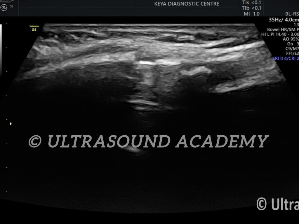
A costochondral fracture is a rare injury involving the cartilage that connects the ribs to the sternum. While X-rays and CT scans are more commonly used for its evaluation, ultrasound can play an adjunctive role, particularly for soft tissue and cartilage injuries. Here is a detailed overview of evaluating a costochondral fracture using ultrasound:
Indications for Ultrasound
When there is localized pain, tenderness, or swelling over the costochondral junction.
Suspected costochondral injury not visible on X-rays (cartilage is radiolucent).
When radiation exposure is to be minimized, e.g., in children or pregnant individuals.
Advantages of Ultrasound
Real-time imaging: Allows assessment of cartilage and adjacent soft tissues.
High-resolution visualization: Effective for detecting disruptions in the cartilage and small hematomas.
Dynamic evaluation: Can detect abnormalities during patient movement or breathing.
No radiation: Safe for repeated use.
Ultrasound Technique
Preparation:
Use a high-frequency linear probe (10–15 MHz) for superficial structures.
Patient is positioned supine or sitting, depending on the location of the suspected fracture.
Properly expose the anterior thoracic wall.
Procedure:
Identify the ribs and their cartilaginous extensions, following their course to the sternum.
Evaluate the costochondral junction bilaterally for comparison.
Perform dynamic maneuvers such as deep breathing or slight compression with the probe to elicit pain and assess structural integrity.
Findings:
Normal appearance: Smooth, continuous cartilage and rib margins.
Fracture: Disruption or irregularity in the echotexture of the costochondral cartilage.
Hematoma: Anechoic or hypoechoic fluid collection adjacent to the cartilage.
Edema: Hypoechoic swelling of the surrounding soft tissue.
Limited penetration in obese patients or those with significant chest wall swelling.
Difficult to visualize deep or posterior costochondral injuries.
Operator-dependent technique.
Clinical Integration
Ultrasound is most useful when combined with clinical findings and other imaging modalities:
If ultrasound findings are inconclusive, a CT scan is often warranted for better structural detail.
Ultrasound can be particularly beneficial for follow-up evaluations to monitor healing.
Ultrasound imaging is a valuable tool for evaluating costochondral fractures, especially when initial radiographs are inconclusive. Key ultrasound findings indicative of a costochondral fracture include:
Fracture Line: A visible discontinuity within the costal cartilage, appearing as a hypoechoic (dark) line traversing the cartilage.
Disruption of the Anterior Echogenic Margin: The smooth, bright (echogenic) anterior surface of the cartilage may appear irregular or interrupted at the fracture site.
Step-off Deformity: A misalignment or displacement at the fracture site, presenting as a discontinuity or “step” in the cartilage contour.
Gas at the Costochondral Junction: The presence of hyperechoic (bright) foci with posterior acoustic shadowing, indicating gas bubbles near the fracture, which can occur due to injury.
Dynamic Instability: Real-time ultrasound may reveal abnormal movement at the fracture site during respiration or gentle palpation, suggesting instability.
It’s important to note that ultrasound is operator-dependent and may have limitations in visualizing deep or posterior costal cartilage injuries. Additionally, distinguishing between calcified cartilage and fractures can be challenging. Therefore, correlating ultrasound findings with clinical assessment and, if necessary, other imaging modalities like CT or MRI is essential for accurate diagnosis.
For a comprehensive understanding of costal cartilage injuries and their imaging characteristics, including ultrasound findings, you may refer to the following sources:
Radiopaedia.org: Provides detailed articles on costal cartilage injuries, including imaging features and differential diagnoses.
Radsource: Offers case studies and imaging examples related to costal cartilage injuries, emphasizing MRI findings but also discussing ultrasound.
BMJ Case Reports: Presents case reports on operative treatment of costochondral dislocations, discussing imaging findings and management strategies.
These resources provide in-depth information on the imaging and management of costochondral fractures.