Ca Buccal mucosa with submandibular gland metastasis
55 years old male referred for ultrasound neck by surgeon in view of swelling over left submandibular region.
Findings and discussion:
On B mode ultrasound:
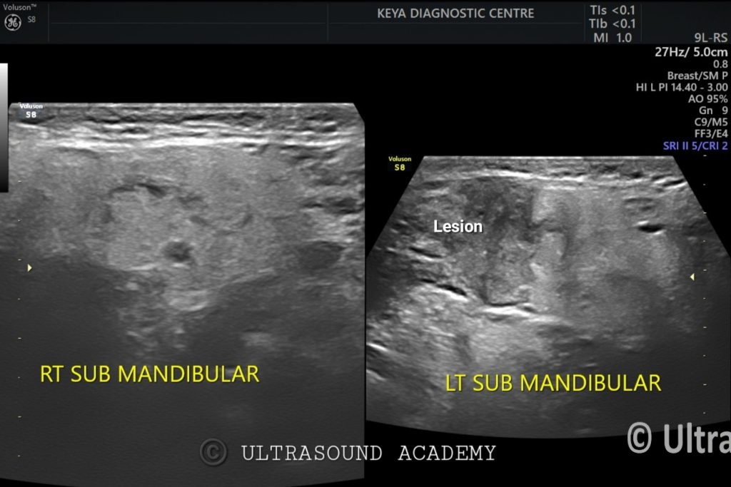
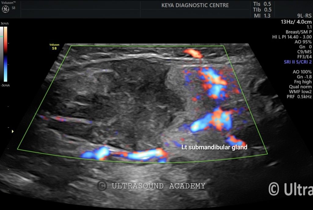
There is an ill-defined heterogenous predominantly hypoechoic lesion is seen involving the left submandibular gland showing vascularity on Color Doppler examination.
On dedicated screening of the entire neck region, the left buccal region showed an irregular ill-defined solid hypoechoic mass showing intralesional and perilesional vascularity on Color Doppler. Also, cortical irregularity is noted.
Right submandibular, bilateral parotid and thyroid gland appears normal.
So, the diagnosis of Ca left buccal mucosa with ipsilateral submandibular gland metastasis is given.
Later confirmation of the diagnosis done by FNAC from left submandibular gland which showed Malignant squamous cells suggesting keratinizing squamous carcinoma.
Ultrasound is not the primary imaging modality for buccal mucosa carcinoma. However, it can be useful in evaluating tumor invasion, lymph node involvement, and vascular structures.
Ultrasound Features of Buccal Mucosa Carcinoma
Hypoechoic Mass: The tumor usually appears as an irregular, hypoechoic lesion compared to surrounding soft tissues.
Loss of Normal Layered Structure: The normal mucosa, submucosa, and muscle layers may appear disrupted or indistinct.
Infiltration into Deeper Tissues: High-resolution ultrasound (especially intraoral ultrasound with a high-frequency probe) can detect muscle invasion (e.g., into the buccinator muscle).
Increased Vascularity: Color Doppler ultrasound may show increased blood flow, indicating tumor angiogenesis.
Lymph Node Involvement: Ultrasound is useful in detecting cervical lymphadenopathy, which may show round, hypoechoic, enlarged nodes with loss of the fatty hilum, suggesting metastatic spread.
Role of Ultrasound in Diagnosis
Intraoral ultrasound (using a high-frequency probe) can be used for detecting tumor depth and invasion.
Neck ultrasound is valuable for assessing regional lymph node metastasis.
Confirmatory Tests
Biopsy is required for definitive diagnosis.
MRI or CT scan provides better soft-tissue contrast and is preferred for staging.