55-year-old male with a 2-month history of a progressively enlarging lesion on the left buccal mucosa. Referred for ultrasound evaluation of tumor extent and regional lymphadenopathy.
Ultrasound Findings and Description:
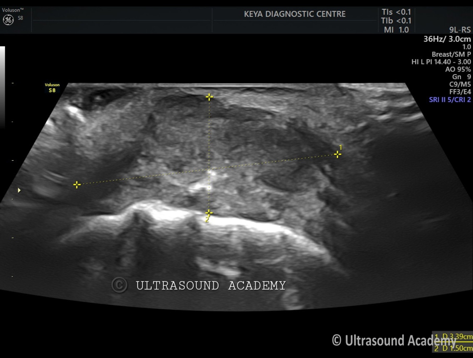
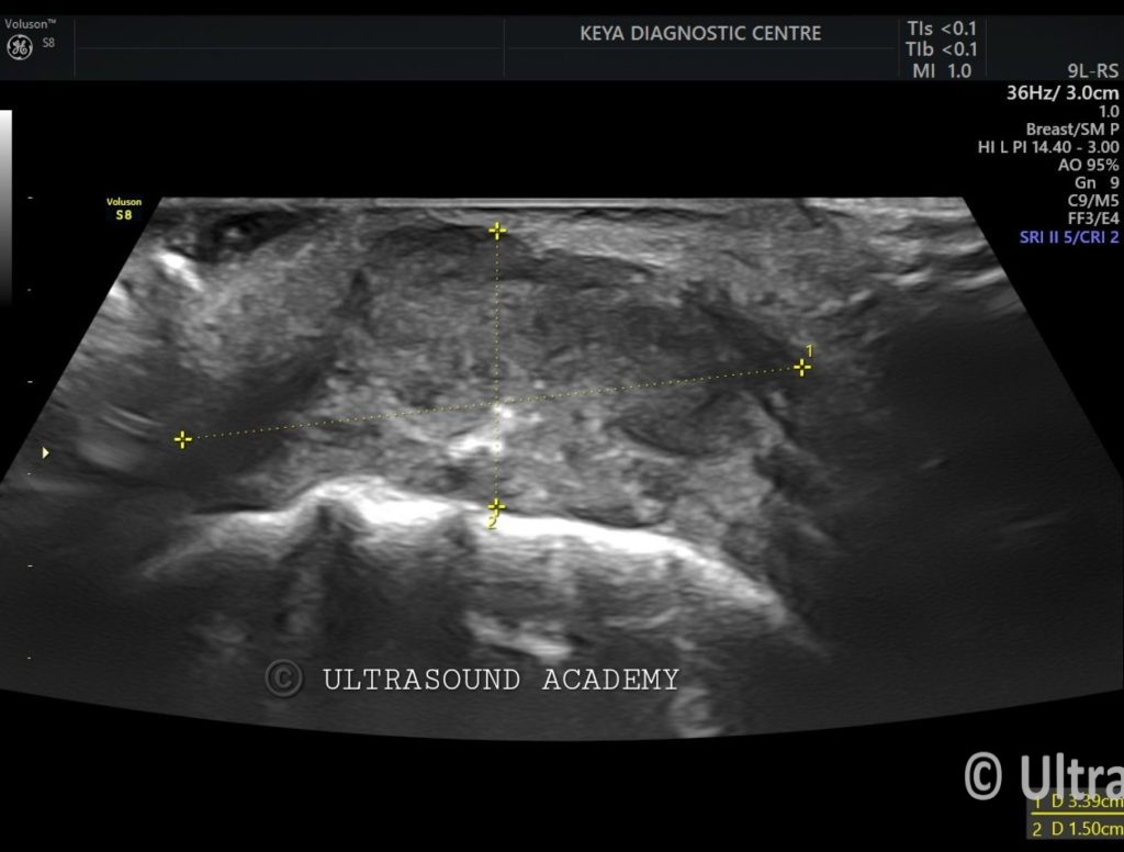
Hypoechoic irregular mass measuring approximately Approx 1.5 cm in greatest dimension located in the left buccal mucosa.
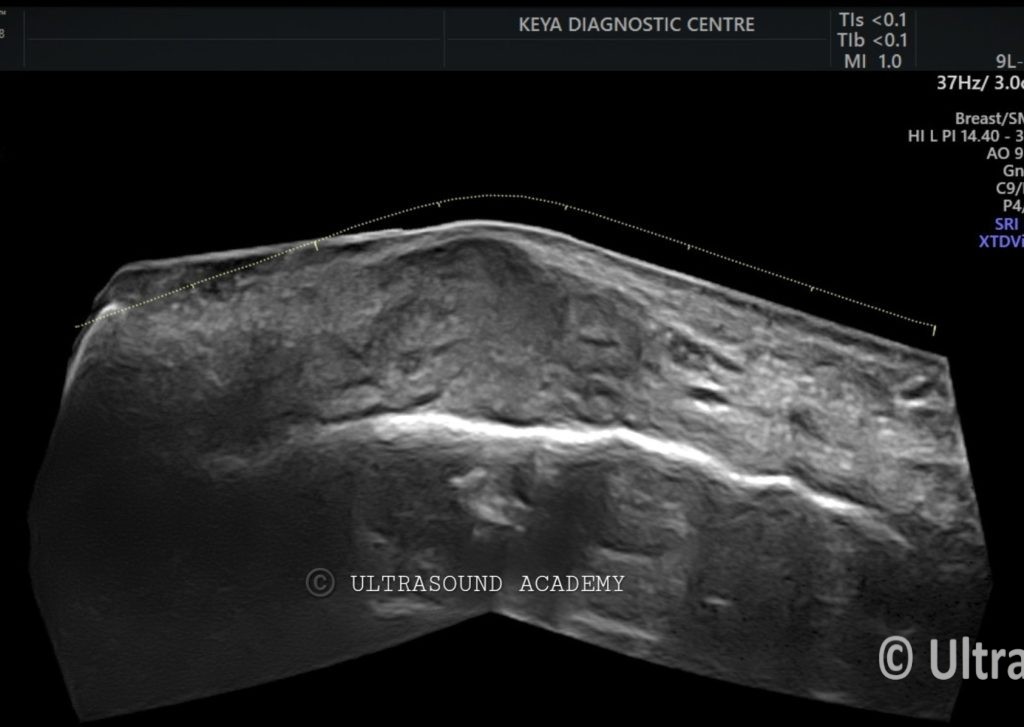
The mass shows heterogeneous echotexture with poorly defined borders, invading into the adjacent soft tissues.
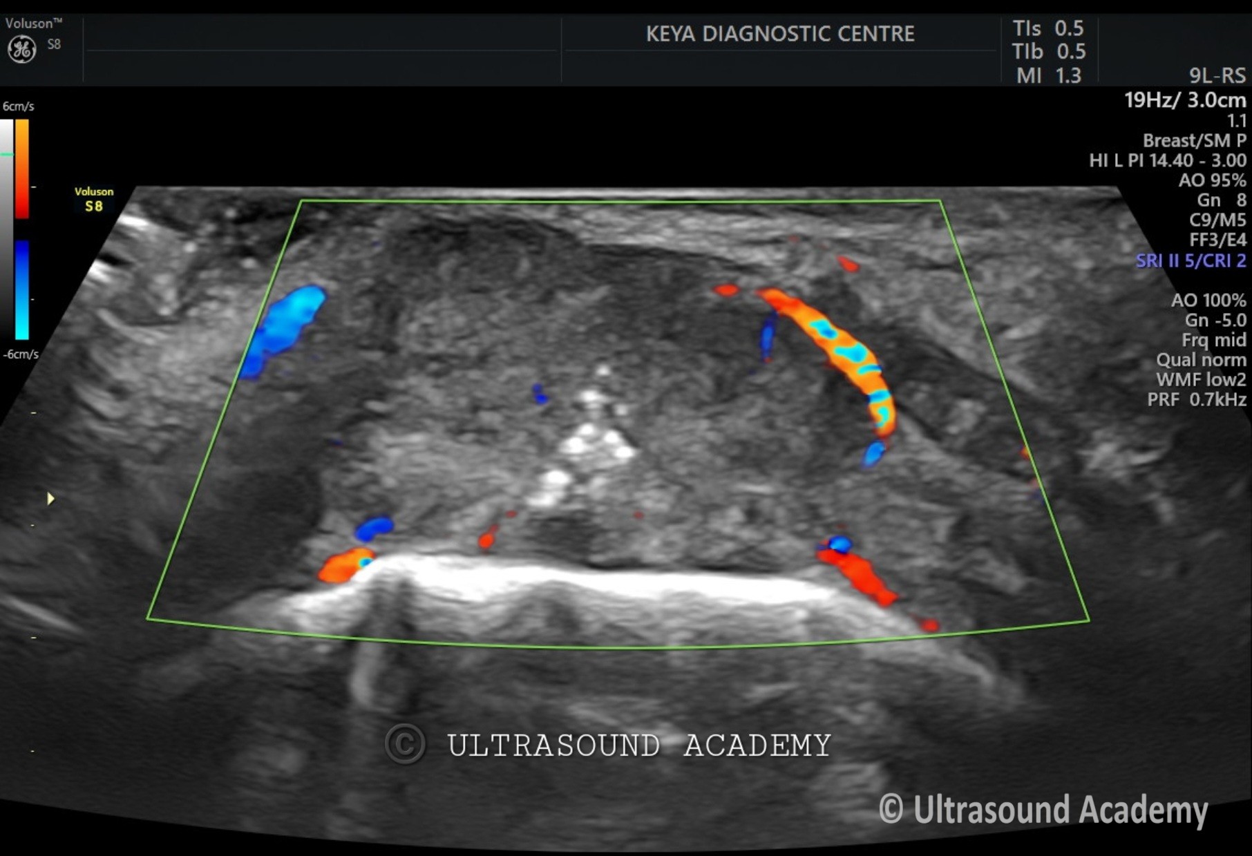
Increased vascularity seen within the lesion on color Doppler.
Also an ulcer with air foci seen as hyperechoic specks.
Rest of the neck was normal. No metastatic LN found in neck.
No evidence of vascular encasement or major vessel involvement.
Biopsy confirmed squamous cell carcinoma.
Diagnosis: Invasive buccal mucosa carcinoma.
Differential Diagnosis:
Mucoepidermoid Carcinoma – Another malignant lesion of salivary origin that may also present as a hypoechoic mass but tends to be more cystic.
Benign Salivary Gland Tumor (e.g., Pleomorphic Adenoma) – Generally well-circumscribed and may appear solid or cystic, unlike the infiltrative appearance seen here.
Discussion:
Buccal mucosa carcinoma, primarily squamous cell carcinoma, is a common form of oral cancer. Ultrasound is useful in evaluating the local extent of the tumor and its relationship to surrounding structures, as well as assessing for lymph node metastasis. In this case, the ultrasound findings suggest advanced local disease with potential regional nodal involvement.
Additional imaging: such as MRI or CT, may be needed to further assess the depth of invasion and distant metastasis.
Conclusions:
Ultrasound findings are consistent with buccal mucosa carcinoma.