History:
A 45-year-old male presents with a painful, swollen left cheek for the past two weeks. The patient reports fever and difficulty chewing. No significant medical history or recent trauma. Referred for an ultrasound of the parotid glands to evaluate the swelling and suspected infection.
Ultrasound Findings :
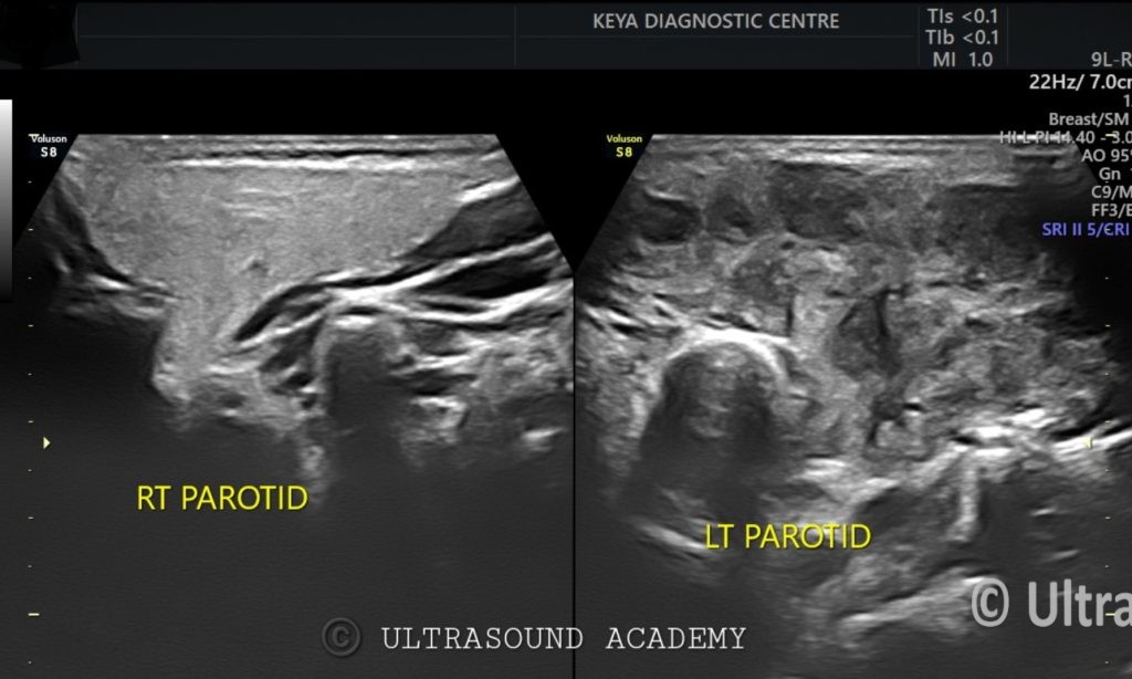
● Right Parotid Gland: Normal in size and echotexture, used as a comparison for the affected side.
● Left Parotid Gland: Diffusely enlarged with multiple hypoechoic lesions scattered throughout the gland, measuring between 1-2 cm. The lesions contain thick internal echoes, suggestive of possible abscess formation.
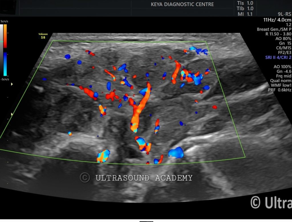
● Color Doppler shows increased internal vascularity, indicating active inflammation within the gland.
● No significant ductal dilation or sialolithiasis observed in either gland.
● Surrounding subcutaneous tissue appears mildly thickened, consistent with local inflammatory changes.
Diagnosis: Acute suppurative parotitis of the left parotid gland with probable abscess formation.
Differential Diagnosis:
1. Chronic Sialadenitis: Long-standing inflammation could lead to gland enlargement and hypoechoic areas, but typically without acute abscess formation.
2. Parotid Gland Tumor (Warthin’s Tumor or Mucoepidermoid Carcinoma): While rare, parotid gland tumors may present with solid and cystic components, but the acute presentation and fever make infection more likely.
3. Sjögren’s Syndrome: Characterized by diffuse glandular enlargement and hypoechoic areas, though abscess formation is uncommon.
Discussion:
● The ultrasound findings are most consistent with acute suppurative parotitis, which is an infection of the parotid gland, often caused by bacterial pathogens like Staphylococcus aureus or Streptococcus.
● The formation of hypoechoic lesions with thick internal echoes likely represents developing abscesses.
● The increased vascularity further supports the diagnosis of acute inflammation.
● The normal appearance of the right parotid gland rules out bilateral involvement.
● Parotitis can occur due to reduced salivary flow, dehydration, or ductal obstruction, leading to bacterial infection.
● Prompt treatment with antibiotics and possible drainage of abscesses is necessary to prevent further complications.
Conclusions:
● Diffuse enlargement of the left parotid gland with hypoechoic lesions and thick echoes, consistent with abscess formation.
● Increased internal vascularity suggests active inflammation, consistent with acute suppurative parotitis.
● Normal right parotid gland for comparison.
● Antibiotic treatment and potential abscess drainage recommended.