


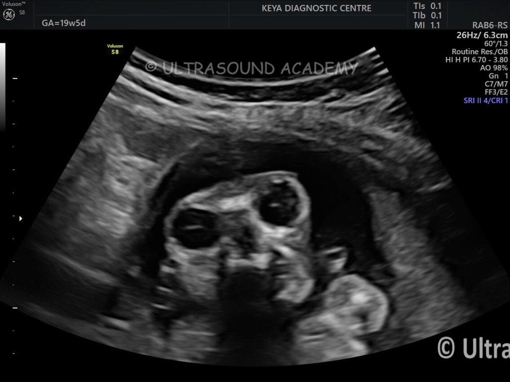

Findings and Discussion:
No brain parenchyma and skull vault bones are seen
Typical ‘ Frog Eye’ appearance is seen due to absence of skull vault and brain parenchyma.
Sometimes some amount of disorganized brain parenchyma is seen then its termed as exencephaly.
Anencephaly is most severe form of neutral tube defect.
It can be diagnosed on USG comfortably as early as 10-11 wks.
MSAFP levels are elevated in open neural tube defects
These major neural tube defects are part of Acrania- Exencephaly- Anencephaly sequel.
Acrania: Congenital disorder characterized by absent/severely malformed skull vault
– Complete or partial absence of cranial bones
Exencephaly:
– Early stage neural tube defect where brain tissue is exposed outside the skull
– Precursor to anencephaly
Anencephaly:
– Severe neural tube defect where major portions of brain, skull, and scalp are missing
– Lethal condition
– Cannot be surgically corrected
Antenatal USG (Ultrasonography) Findings:
– Can be suspected during early pregnancy as early as 9- 10 wks
– Typically diagnosed between 11-14 weeks gestation
– Key markers include absence of skull bones, exposed brain tissue
– Critical for early diagnosis and pregnancy termination
Preventive measures: Folic acid supplementation before and during early pregnancy can help reduce neural tube defect risks.

