3rd or 4th BRANCHIAL CLEFT SINUS – Case By Dr. Nitin Jadhav
History:
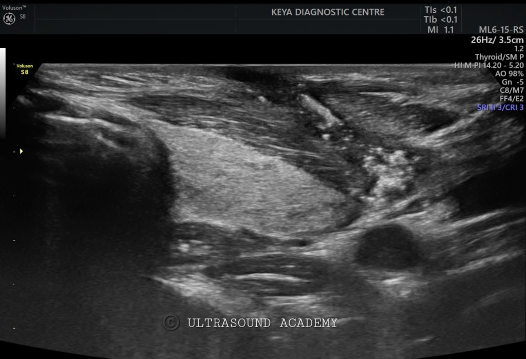
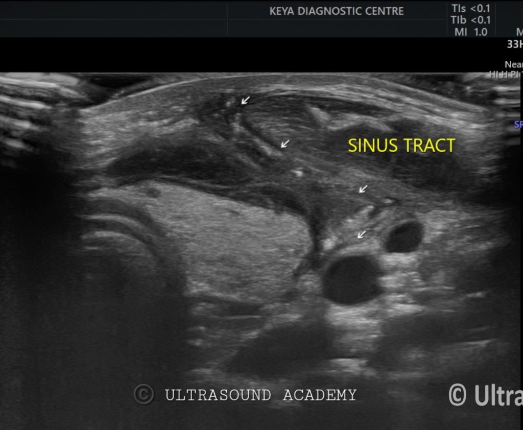
Patient presented with recurrent neck infections or abscesses, located in the lower neck.
Ultrasound Findings and Description:
● Fluid-filled sinus tract is identified near the lower anterior neck, adjacent to the thyroid gland or extending toward the pyriform sinus.
● Increased vascularity in the surrounding tissues may be noted in cases of infection or abscess formation.
● Associated Findings: Possible abscess formation or inflammation surrounding the sinus tract during infection.
● Thyroid Gland: May appear normal or show reactive changes due to proximity to the sinus tract.
Diagnosis: 3rd or 4th branchial cleft sinus
Differential Diagnosis:
1. Thyroglossal Duct Cyst: Midline cystic structure, typically located higher in the neck and moves with swallowing.
2. Branchial Cleft Cyst (2nd branchial cleft): Cystic mass found at the level of the hyoid bone, lateral to the neck, without extension to the pyriform sinus.
3. Lymphadenopathy: Enlarged lymph nodes that may mimic cystic structures but are solid and often hypervascular on Doppler imaging.
Discussion:
● Branchial cleft sinuses are rare congenital anomalies, with the 3rd and 4th branchial clefts being less common than the 2nd. They frequently present with recurrent infections, and imaging is essential for diagnosis. Ultrasound is the first-line modality, though additional imaging such as CT or MRI may be required for surgical planning.
Conclusions:
● Ultrasound is valuable for identifying branchial cleft sinuses, particularly in locating the tract and assessing for associated infection or abscess. Doppler imaging assists in evaluating inflammation and vascularity around the sinus.
error: Content is protected !!