Findings and Discussion:
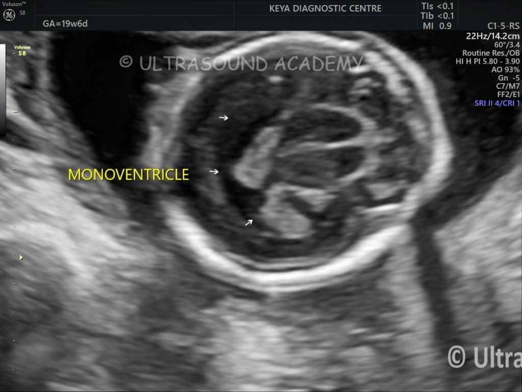
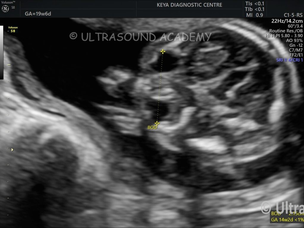
Single monoventricle is seen.
Both thalami appears uncleaved.
Anteriorly interhemispheric fissure is absent.
CSP, corpus callosum appears absent.
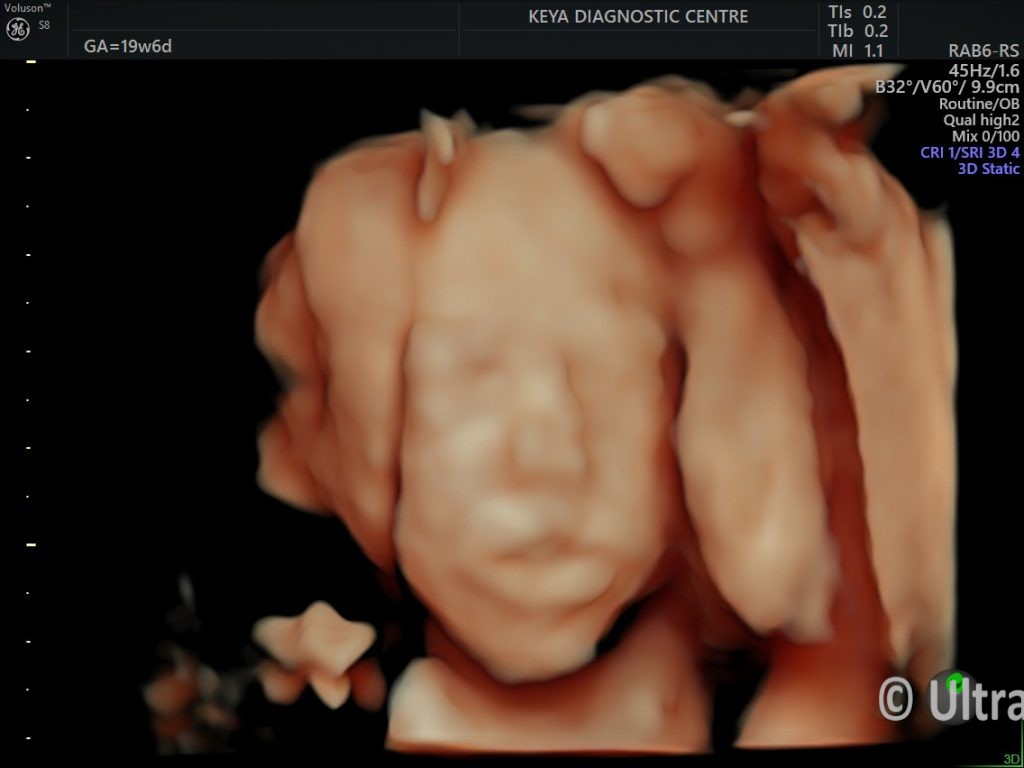
Hypotelorism noted.
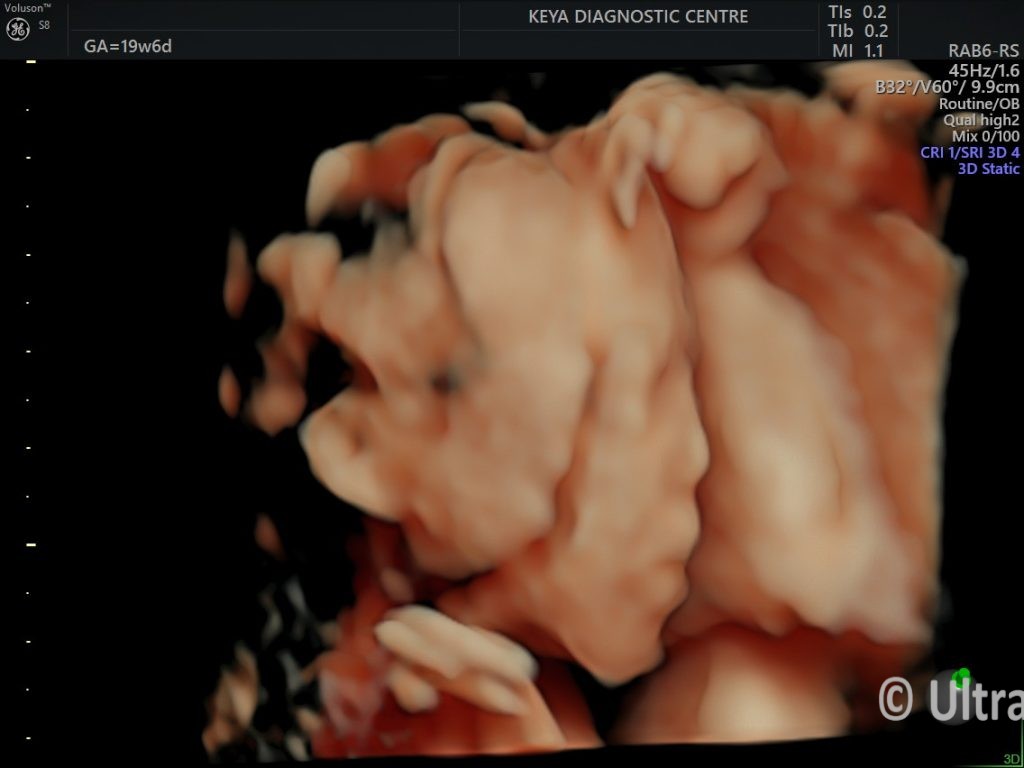
No other facial deformity/ cleft noted.
Pregnancy terminated.
Holoprosencephaly is a rare congenital brain malformation resulting from the incomplete division of the prosencephalon (forebrain) into two hemispheres during early embryonic development. Antenatal ultrasonography (USG) plays a critical role in the diagnosis of this condition.
Antenatal USG Features of Holoprosencephaly:
Holoprosencephaly is classified into four types based on severity, and the findings vary accordingly:
- Alobar Holoprosencephaly (Most Severe):
Single ventricle: Absence of the normal division of the lateral ventricles.
Uncleaved thalami: The thalami appear as a single mass without normal separation.
Absent midline structures: Absence of the falx cerebri, corpus callosum, and interhemispheric fissure.
Monoventricle appearance: A large single central fluid-filled cavity.
Facial anomalies: Associated facial abnormalities like cyclopia, proboscis, hypotelorism, or cleft lip/palate may be detected.
- Semilobar Holoprosencephaly (Intermediate Severity):
Partial separation of hemispheres: Some division of the brain is noted posteriorly.
Uncleaved structures: The anterior portions of the brain, including the thalami, may remain uncleaved.
Abnormal ventricular configuration: An enlarged single ventricle with partial septation.
Facial abnormalities: Similar to alobar but may be milder.
- Lobar Holoprosencephaly (Mildest Form):
Near-complete separation of the cerebral hemispheres, with abnormal non cleavage of structures only in the frontal region.
Normal or mildly dysplastic ventricles: Ventricles may appear relatively normal but with evidence of midline defects.
May lack overt facial abnormalities.
- Middle interhemispheric Variant (MIHV)-
This is a rare type of Holoprosencephaly where there is cleavage at anterior and posterior part of brain with non cleavage seen in mid part of brain.
There may fusion of lateral ventricles at parietal regions.
Facial abnormalities may not be seen.
Facial Anomalies Commonly Associated with Holoprosencephaly
Cyclopia (single orbit).
Hypotelorism (closely spaced eyes).
Midline cleft lip/palate.
Proboscis (tubular nasal structure).
Absent nasal bone.
Key Sonographic Findings:
Transabdominal and Transvaginal Views: Transvaginal USG may provide better resolution, especially in early gestation.
Midline Abnormalities: Evaluate the presence of the falx cerebri, interhemispheric fissure, and cavum septum pellucidum.
Craniofacial Abnormalities: Assess the orbits, nose, and maxillary profile for characteristic facial defects.
Hydrocephalus or Microcephaly: Secondary brain abnormalities may be present.
Polyhydramnios: Frequently associated due to swallowing difficulties.
Timing of Diagnosis:
Holoprosencephaly can typically be diagnosed as early as 11–13 weeks of gestation during the first-trimester scan using transvaginal ultrasound.
Advanced second- and third-trimester scans can confirm and classify the type.
Role of Additional Imaging:
MRI: Fetal MRI can complement USG, especially for a detailed evaluation of the brain and midline structures.
3D Ultrasound: May provide enhanced visualization of craniofacial anomalies.
Early diagnosis is crucial for counseling regarding prognosis, as severe forms (alobar and semilobar) are often associated with poor outcomes. Genetic counseling and evaluation for chromosomal abnormalities (e.g., Trisomy 13) are also advised.