ANGULAR PREGNANCY 2 – Case By Dr. Nitin Jadhav
History:
Patient presents with early pregnancy, reporting pelvic pain and mild vaginal bleeding.
Findings and Description:
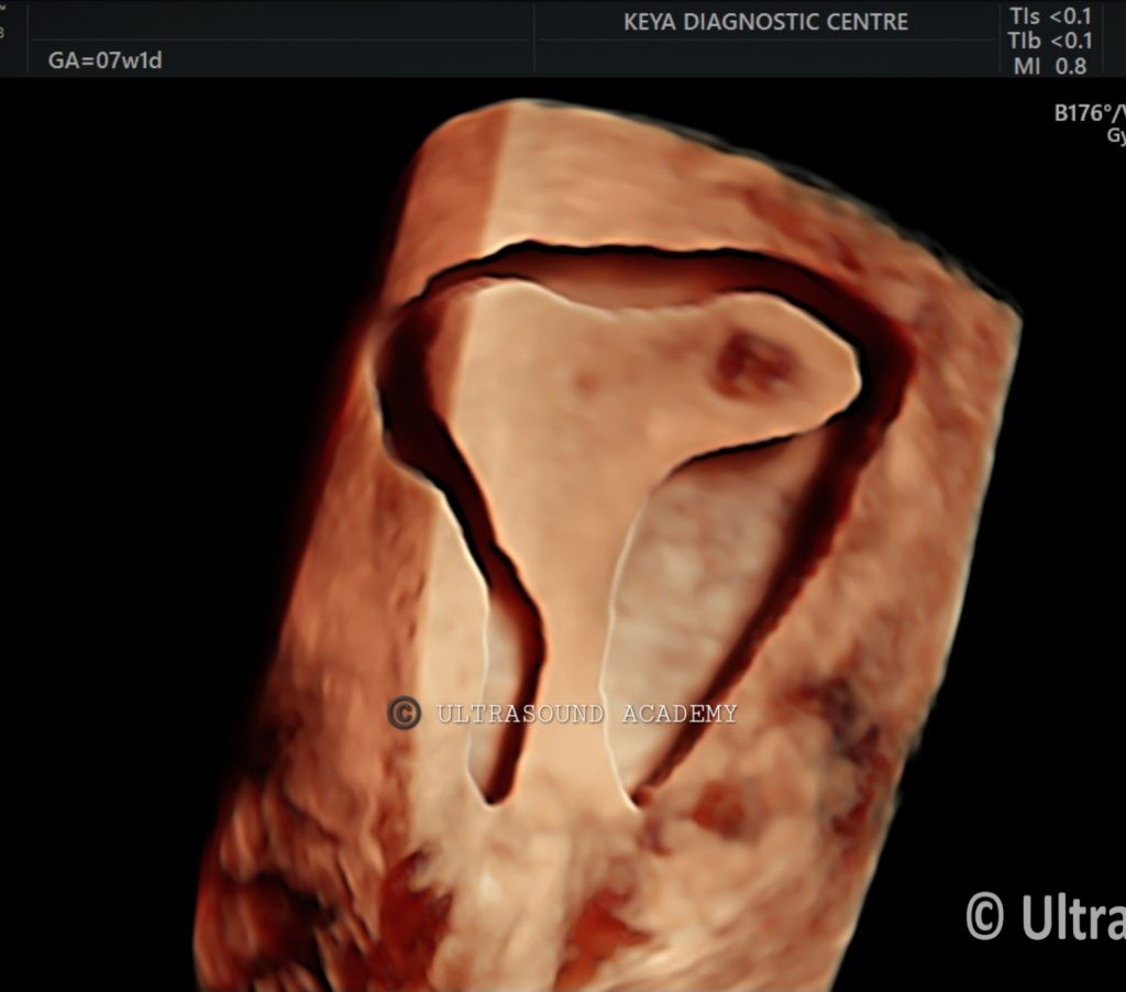
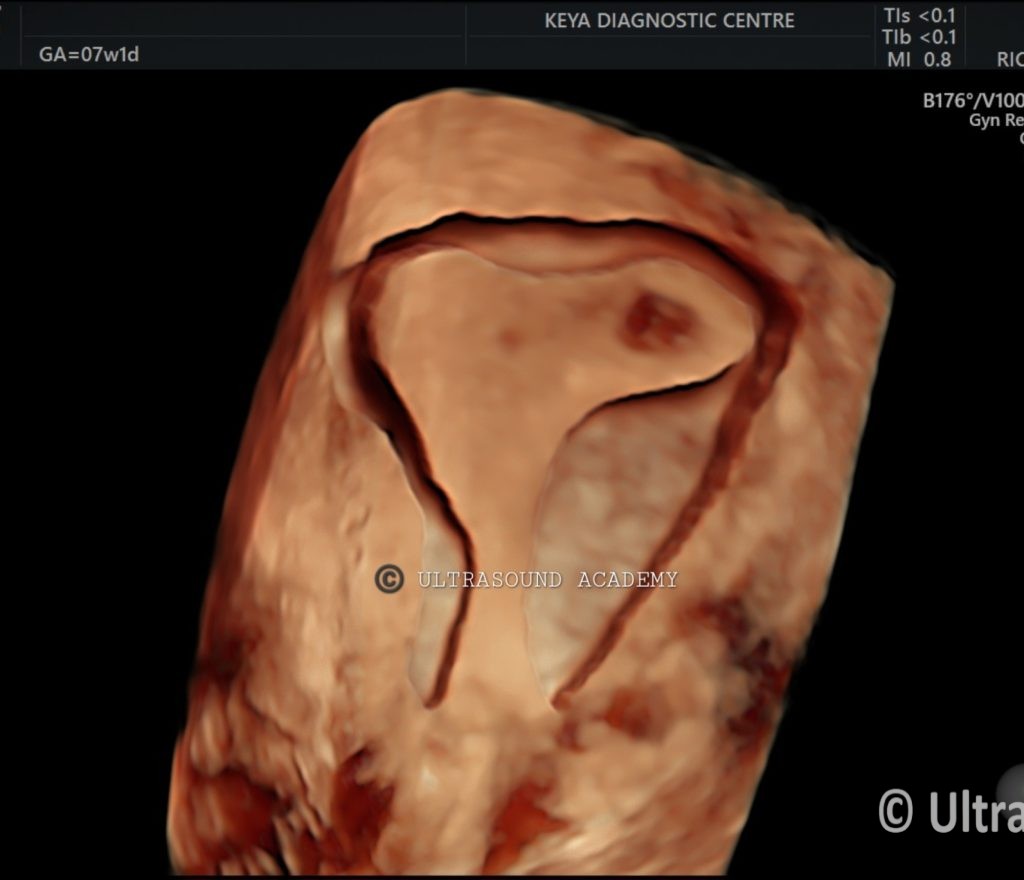
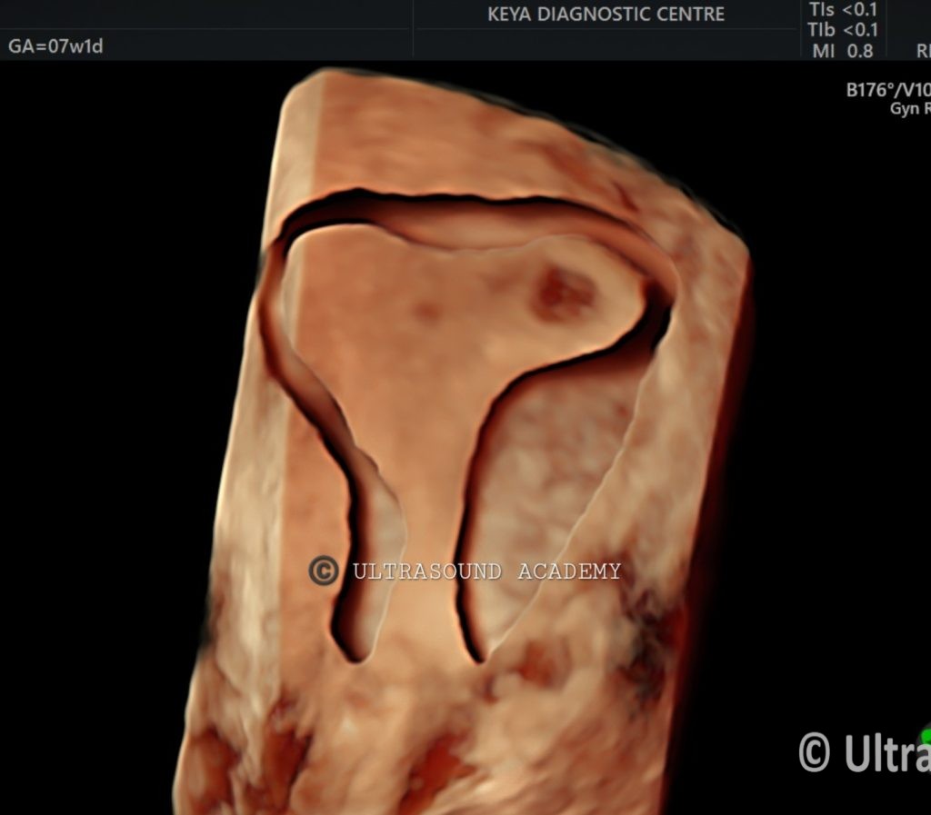
- This is an 3D transvaginal ultrasound,Findings that may suggest an angular pregnancy include,the gestational sac is located eccentrically within one of the uterine horns, close to the uterine corner but still within the endometrium.
- Gestational sac appears distorted, with irregular shape and proximity to the uterotubal junction, suggesting an angular pregnancy.
- Adequate myometrial thickness seen surrounding the gestational sac, differentiating this from an ectopic or interstitial pregnancy.
- 3D ultrasound is particularly helpful in these cases because it provides better spatial orientation and visualization of the uterus and sac compared to traditional 2D ultrasound. Early detection through imaging is crucial in managing the condition and preventing complications.
- Fetal pole and heartbeat may be visualized on TVS, but viability can be compromised depending on the extent of implantation near the cornual region.
Diagnosis: Angular Pregnancy / Eccentrically located pregnancy
New updates:Regarding the consensus (not evidence-based) made by
The European Society of Human Reproduction and Embry-
ology or ESHRE Working Group on Ectopic Pregnancy in
2020, the term “angular pregnancy” was proposed to be
abandoned, and “eccentric pregnancy” was recommended by
other colleagues.However, in the literature published after
2020, “angular pregnancy” is still the most commonly used
and an acceptable term for this condition.
Differential Diagnosis:
1 Interstitial Pregnancy: A more dangerous type of ectopic pregnancy where the gestational sac implants within the muscular portion of the fallopian tube. This is often confused with angular pregnancy, but the sac is located more laterally, outside the uterine cavity.
2. Cornual Pregnancy: A type of ectopic pregnancy that occurs in the upper and lateral part of the uterus, which can be mistaken for an angular pregnancy but is more likely to rupture and carries higher risk.
Discussion:
● Angular pregnancy occurs when implantation is near the uterotubal junction but within the uterine cavity. It differs from interstitial or cornual pregnancies, which are outside the main uterine cavity and carry higher risks of rupture.
● Risks associated with angular pregnancy include miscarriage, uterine rupture, and potential complications during delivery. Close monitoring with serial ultrasounds is crucial to assess fetal development and the risk of complications.
● Management depends on the progression of the pregnancy. If the pregnancy is viable, careful monitoring is advised; otherwise, intervention may be necessary to avoid complications such as uterine rupture.
Conclusions:
● Angular pregnancy confirmed by transvaginal ultrasound, with the gestational sac positioned near the uterotubal junction but within the uterine cavity.
● Close monitoring is essential due to the risk of miscarriage and uterine rupture.
● Delivery planning should include considerations for potential complications in the third trimester or at the time of delivery.
error: Content is protected !!