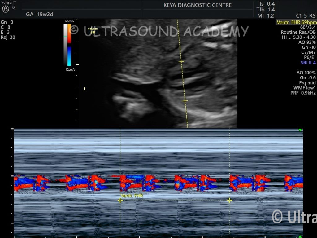
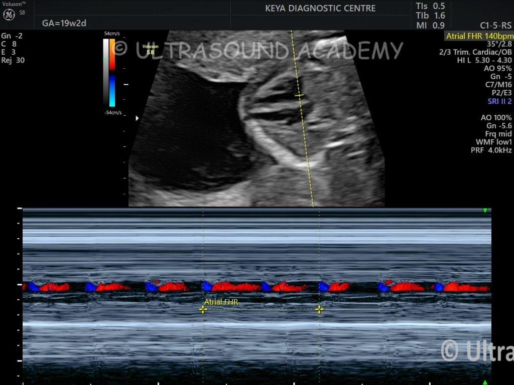
Findings and Discussion- Images from 19 wks scan of Primigravida patient.
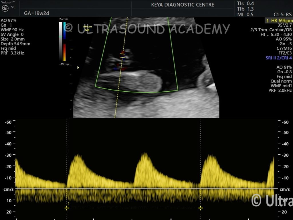
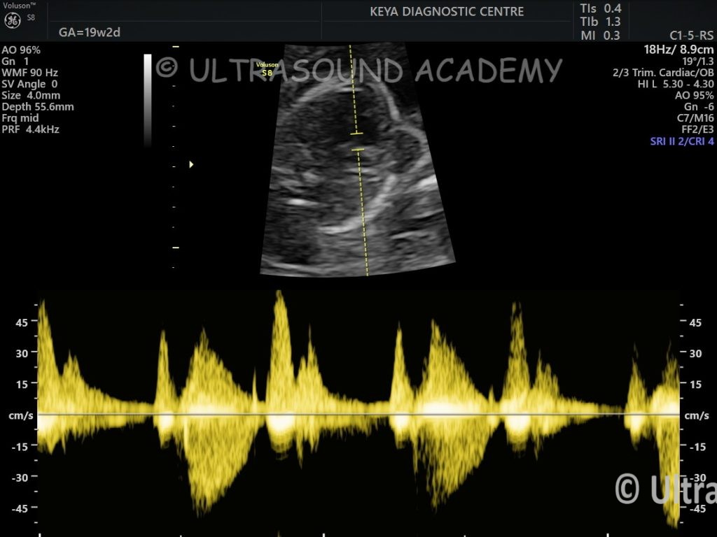
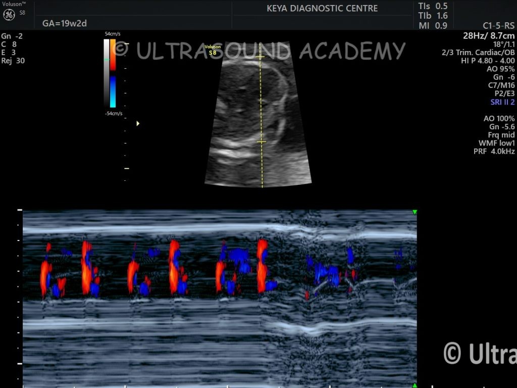
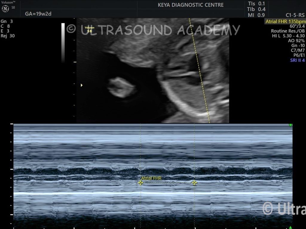
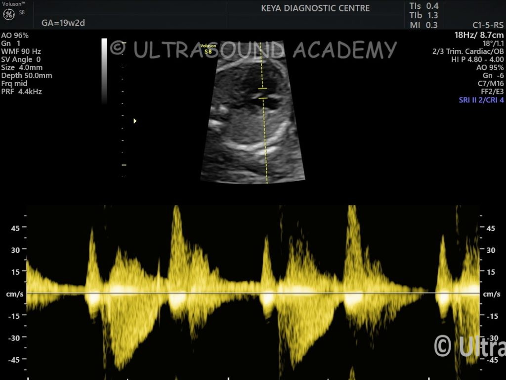
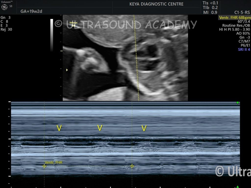
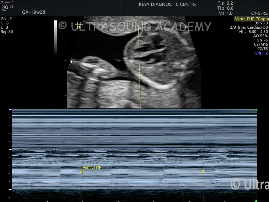
Fetal abnormal cardiac rhythm is noted.
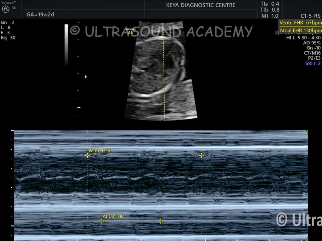
On M mode it is clear that ventricular heart rate is 70 bpm and atrial heart rate is 140 bpm.
For every alternate atrial beat ventricular beat is missing.
These findings confirmed 2:1 AV block.
It was seen intermittently. This 2:1 AV block was seen intermittently for almost 2-3 wks.
Patient was evaluated for autoimmune profile which turned out to be normal.
At 28 wks scan AV block normalised completely.
AV block completely disappeared on all further follow up scan.
It was transient 2:1 AV block.
Detection of Cardiac Arrhythmia
Fetal arrhythmias can be detected during routine antenatal USG, typically when the fetal heart rate is irregular, too fast (tachycardia), or too slow (bradycardia). Advanced imaging techniques such as M-mode ultrasound or Doppler ultrasound are used for detailed assessment.
Types of Fetal Cardiac Arrhythmias
Irregular rhythms:
Most commonly caused by premature atrial contractions (PACs).
Atrial flutter: Organized atrial rhythm with a rate of 300–500 bpm.
Persistent tachyarrhythmias can lead to complications like fetal hydrops.
Bradyarrhythmias:
Sinus bradycardia: Heart rate <110 bpm.
Complete heart block (CHB): Associated with congenital structural abnormalities or maternal autoimmune diseases (e.g., anti-Ro/SSA antibodies).
Assessment Techniques
M-Mode Ultrasound:
Displays the motion of the heart walls and valves over time.
Used to measure atrial and ventricular contraction rates.
Doppler Ultrasound:
Simultaneously measures atrial and ventricular contraction rates to determine the relationship between them.
Fetal Echocardiography:
Essential for diagnosing structural anomalies or more complex rhythm disturbances.
Management
Monitoring: Most arrhythmias, particularly PACs, are benign and require only follow-up.
Medications: For sustained tachyarrhythmias or bradyarrhythmias causing complications, transplacental antiarrhythmic drugs (e.g., digoxin, sotalol) may be administered to the mother.
Delivery planning: Severe arrhythmias or associated complications (e.g., hydrops fetalis) may necessitate delivery at a tertiary care center with neonatal cardiac expertise.