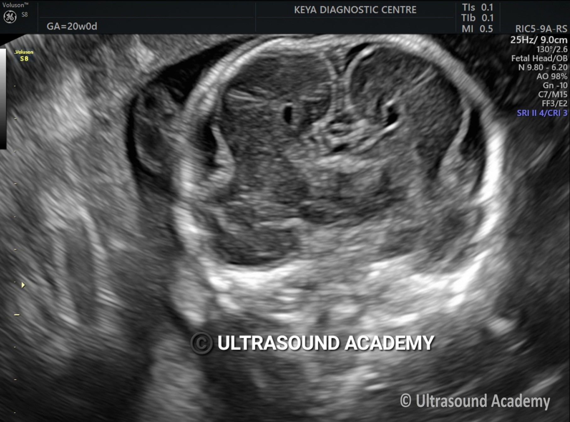
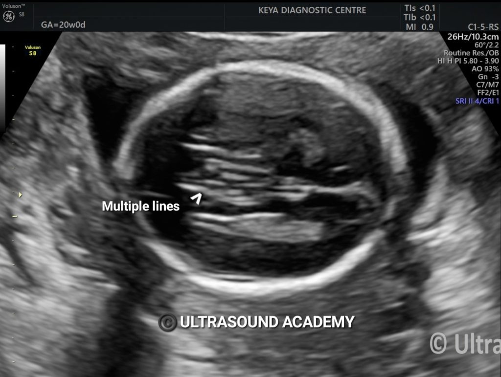
Findings : Absence of corpus callosum. Frontal horns appears widely separated, Absence of CSP.
Corpus callosum agenesis :
It results of an insult occur at approx. 8-12 weeks of gestation.
Incidence rate : 1 : 1000
It may be complete (agenesis) or partial (dysgenesis). When complete, the lateral ventricles displaced laterally and there is upward displacement of third ventricle.
It divided into primary or secondary agenesis.
· Primary agenesis : corpus callosum never forms.
· Secondary dysgenesis : corpus callosum forms normally and is subsequently destroyed.
Associate anamolies includes : Aneuploidy like Trisomy 18,13 and 8. Other association includes : Apert syndrome, Fetal alcohol syndrome, Fryns syndrome, Lowe syndrome, Chiari II malformation, Dandy-Walker spectrum, Holoprosencephaly, Porencephaly, Schizencephaly, Mucopolysaccharidoses, etc…The anamoly is associated with a lipoma of a corpus callosum with variety of developmental dyscrasias including absence of septum pellucidum.
ACC can be identified prenatally on ultrasound, after 18 weeks’ gestation when the CC assumes its final shape. However, ACC can be suspected earlier as parts of the CC can already be identified with high resolution probes and a transvaginal approach. Recently, diagnosis of the CC has been demonstrated by fetal ultrasound as early as 13 weeks and nomograms for its length have been devised. Additionally, the use of the pericallosal artery in the first trimester as an indirect marker of normal CC development has been previously studied and validated.
Ultrasound findings on ultrasound :
· third ventricle 5
o dilated
o can be elevated or dorsally displaced 8
o may communicate with the interhemispheric cistern
o may project superiorly as a dorsal cyst
o choroid may be seen as an echogenic structure in the roof of the cyst
· lateral ventricles
o widely spaced parallel bodies (racing car sign)
o small frontal horns
o colpocephaly: which can give a “teardrop” configuration on axial scans
· septum pellucidum: absent
· interhemispheric fissures: widened
· gyri: may be seen in a “sunray appearance” on the sagittal plane
colour Doppler study may show an abnormal course of pericallosal arteries