CHOLEDOCHOLITHIASIS

USG Findings: Multiple echogenic calculi of varying sizes seen in dilated common bile duct.

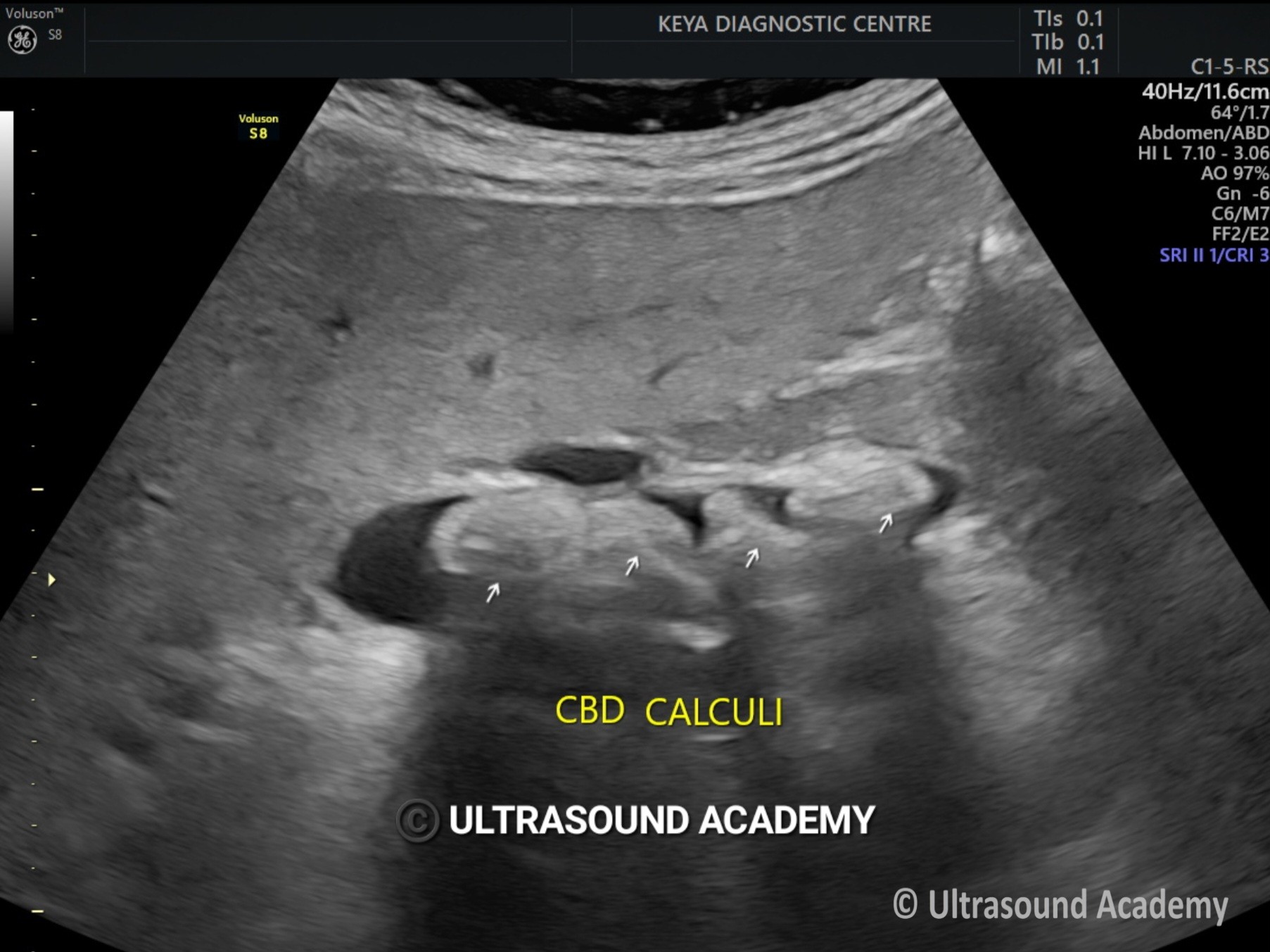
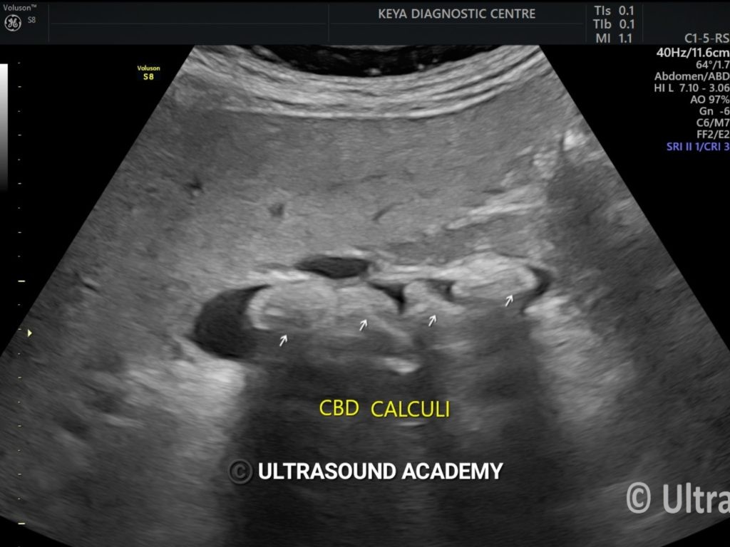
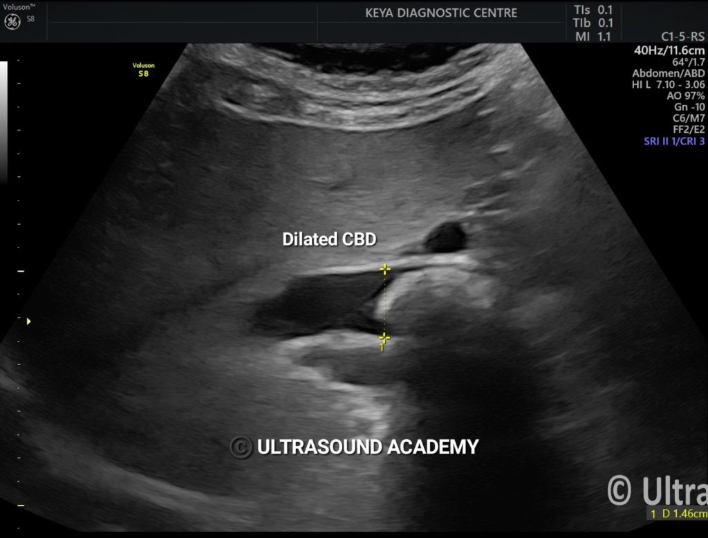
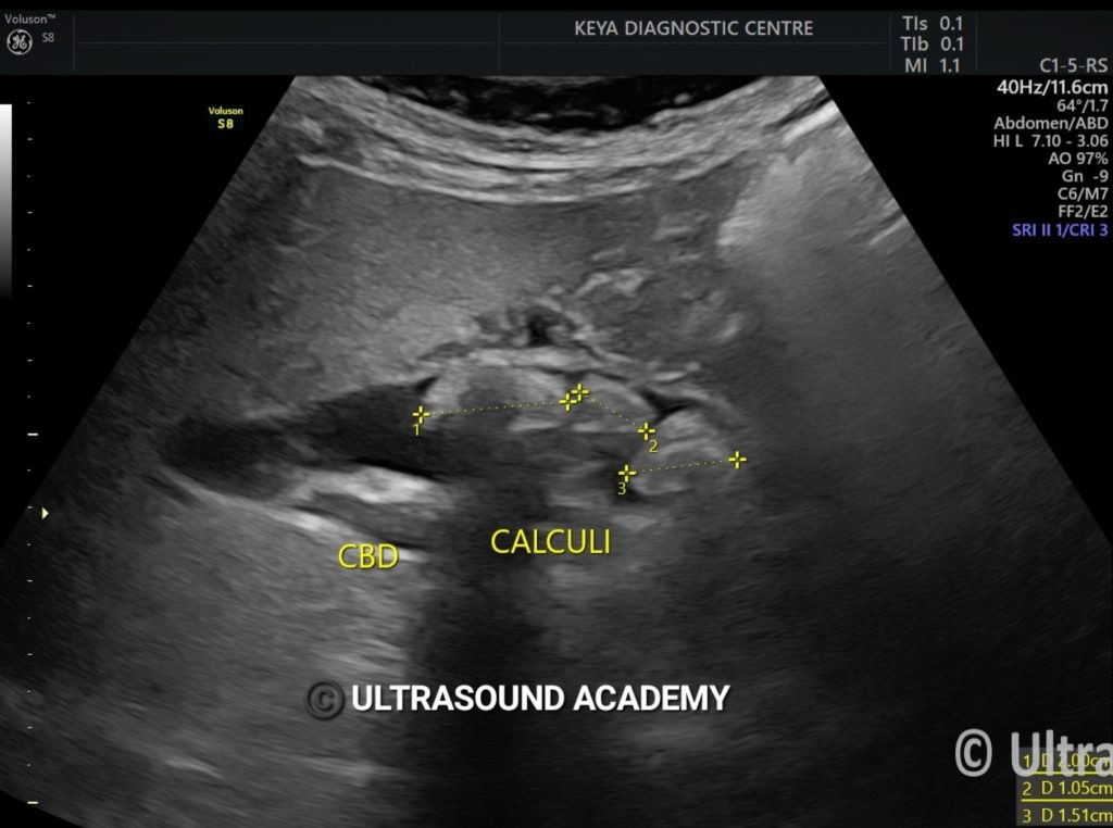
CBD is significantly dilated.

Mild central IHBR dilatation is also seen.

Post cholecystectomy status noted.


Choledocholithiasis refers to the presence of gallstones in the common bile duct (CBD). Ultrasound  is a commonly used imaging technique to detect it.

Ultrasound Findings of Choledocholithiasis:

  1. Dilated Common Bile Duct (CBD)
    • Normal CBD diameter:
      • <6 mm in young adults
      • Up to 8 mm in elderly
      • Up to 10 mm post-cholecystectomy
    • A CBD >7-8 mm in a patient with gallstones suggests obstruction.
  1. Echogenic (Hyperechoic) Foci with Posterior Acoustic Shadowing
    • Stones appear as bright structures with shadows.
    • Sometimes, small stones may not cast shadows.
  1. Floating or Impacted Stones
    • Stones can be mobile or fixed in the CBD.
  1. Sludge or Debris in the CBD
    • Seen as low-level internal echoes without shadowing.
  1. Intrahepatic Bile Duct Dilatation
    • If severe, biliary tree upstream of obstruction expands.

Limitations of USG

  • Small CBD stones (<5 mm) may not be seen.
  • Gas in bowel can obscure visualization.
  • Obese patients may have limited USG penetration.

Next Steps if USG is Inconclusive

  • MRCP (Magnetic Resonance Cholangiopancreatography): More sensitive for detecting stones.
  • ERCP (Endoscopic Retrograde Cholangiopancreatography): Diagnostic & therapeutic (stone removal).
  • CT Scan: Can detect calcified stones but is less sensitive than MRCP

 Further Reading:

                                                                                                                                                      5

image

error: Content is protected !!