Elderly female with a history of abnormal vaginal bleeding and pelvic pain.
Referred for pelvic ultrasound to assess for mass or cervical abnormality.
Ultrasound Findings and Description:
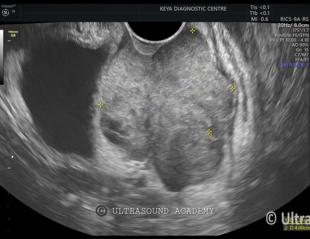
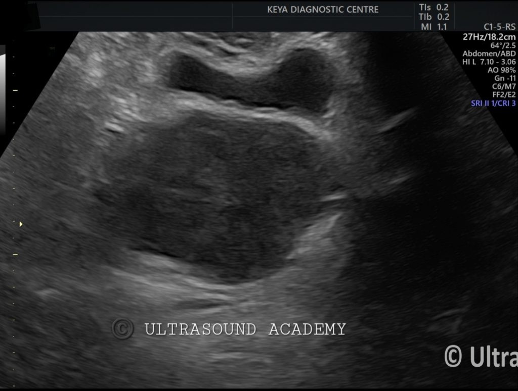
Enlarged cervix with a hypoechoic, irregular mass measuring 5.2 X 4.0 cm, involving the cervical canal and extending into the parametrium.
The mass shows heterogeneous echotexture.
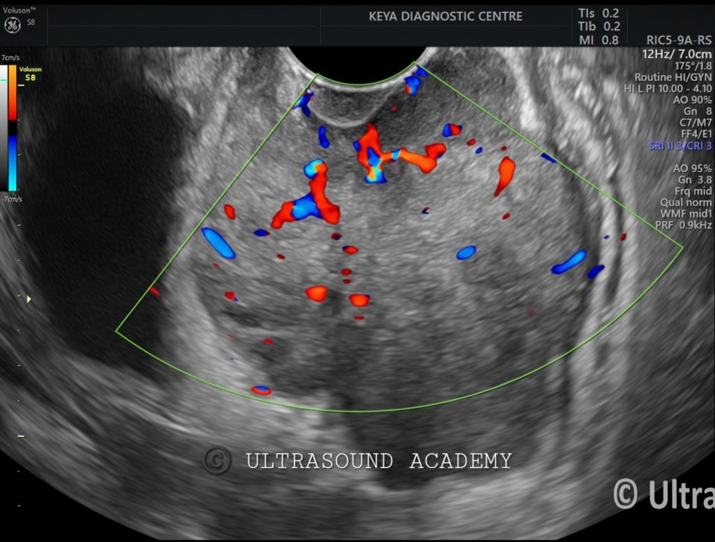
Increased vascularity on Doppler imaging.
Also in such cases look for
endometrial stripe, invasion into the uterine body.
Bilateral adnexal involvement and any free fluid or signs of hydronephrosis.
Diagnosis: Carcinoma of the cervix.
Differential Diagnosis:
Cervical Adenocarcinoma – Typically presents similarly to squamous carcinoma, though may appear more heterogeneous.
Cervical Fibroid (Leiomyoma) – Benign smooth muscle tumor that may appear as a hypoechoic or heterogeneous mass, but typically with well-defined borders.
Discussion:
Cervical carcinoma, especially squamous cell carcinoma, is a common malignancy in women, often linked to human papillomavirus (HPV) infection. Ultrasound is helpful for detecting mass lesions, assessing local spread, and identifying any hydronephrosis from ureteral obstruction.
However, MRI is generally preferred for evaluating the depth of tumor invasion and for staging purposes. In this case, the ultrasound suggests advanced local disease, with parametrium involvement.
Conclusions:
Ultrasound findings are consistent with cervical carcinoma, with probable parametrial invasion. Further imaging with MRI or CT is recommended for detailed staging.