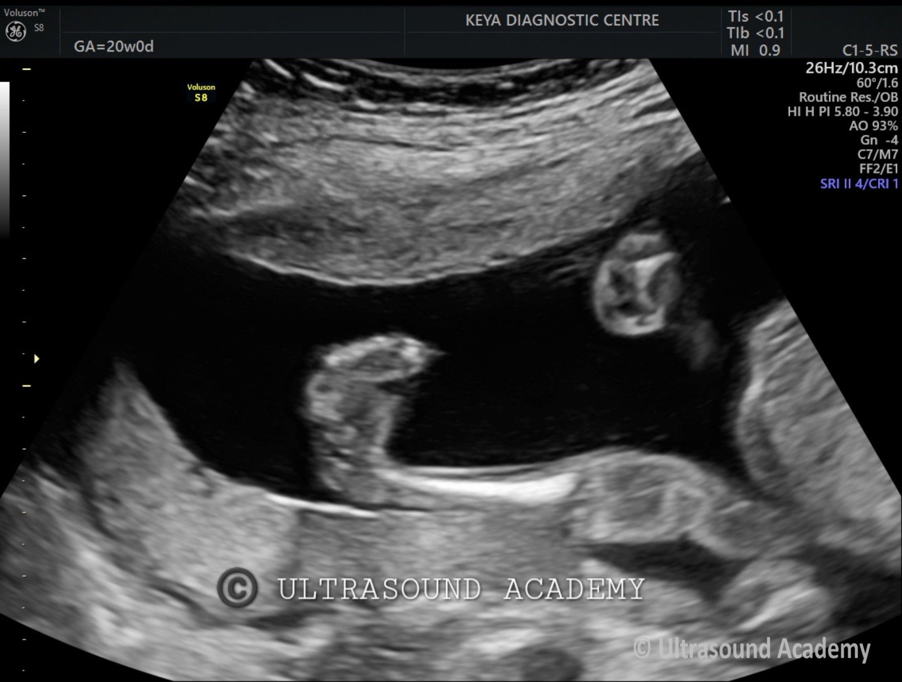
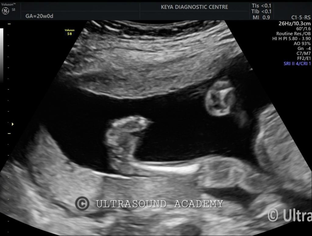
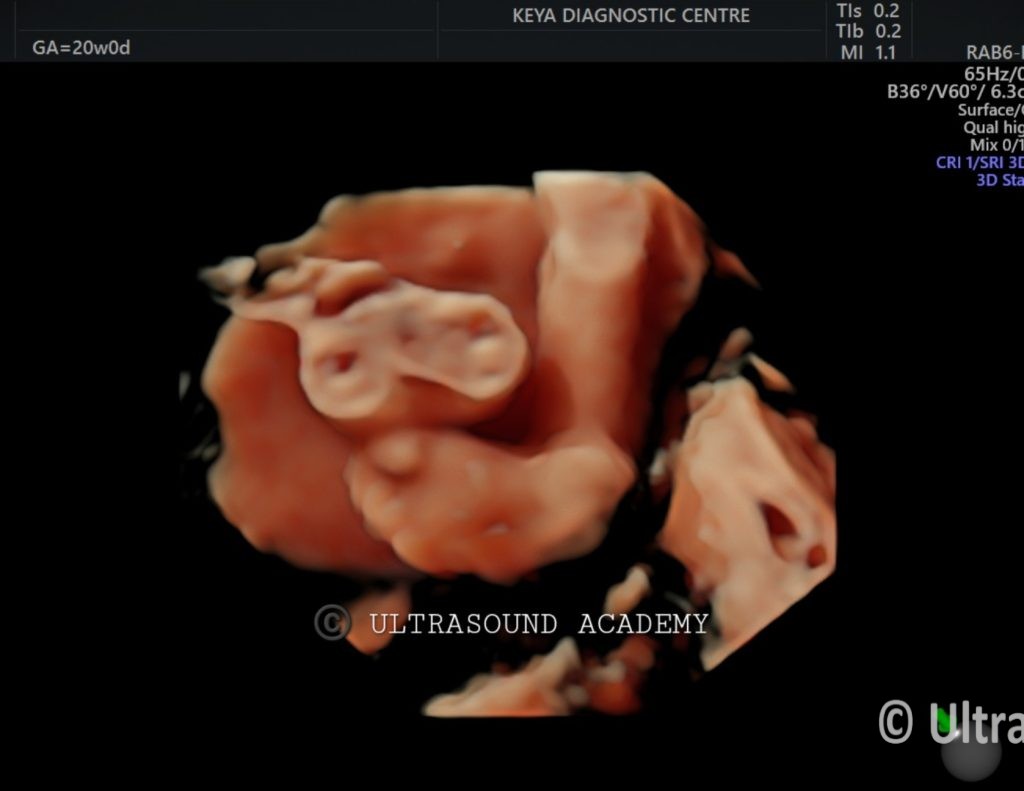
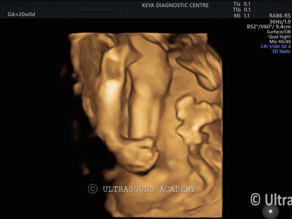
Positional Clubfoot: Feet appear turned inward but without structural bone abnormalities, usually due to intrauterine compression, and resolve spontaneously after birth.
Syndromic CTEV (e.g., Spina Bifida): Clubfoot associated with neurological or spinal abnormalities, such as spina bifida, which may present with other anomalies on ultrasound.
Arthrogryposis Multiplex Congenita: Characterized by joint contractures, including clubfoot, along with abnormalities of other limbs.
Discussion:
Bilateral congenital talipes equinovarus is a common congenital anomaly and can occur as an isolated condition or be associated with other congenital syndromes. Early detection via ultrasound allows for parental counseling and consideration of postnatal treatment options, which typically involve serial casting or corrective surgery.
Conclusions:
Ultrasound findings of inward foot rotation, restricted ankle movement, and abnormal bone orientation are characteristic of bilateral congenital talipes equinovarus. Prenatal identification enables early intervention to achieve functional correction after birth.