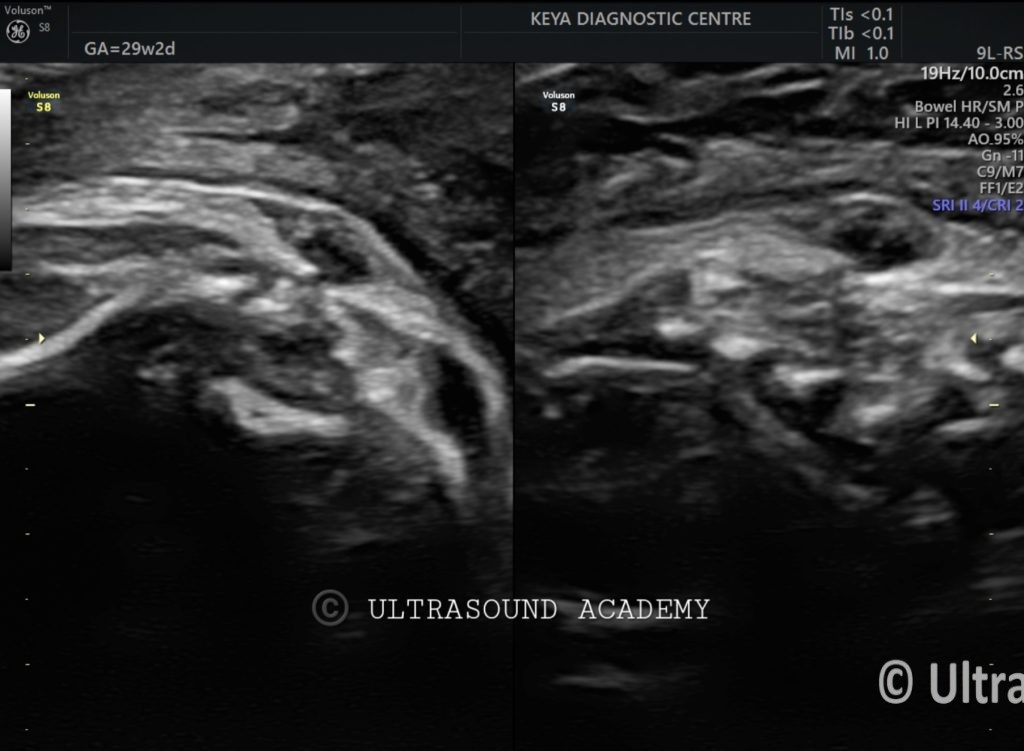
30 years old female referred by obstetrician for routine growth scan at 29 weeks of gestation.
Findings and discussion:
On B mode ultrasound image and cine: (Axial / coronal / sagittal sections)
There is small, well-defined fluid filled anechoic cystic lesion is seen at lower lumbar spinal level.
No internal echoes noted.
The lesion is seen communicating with the neural canal.
The overlying skin appears to be intact.
No evidence of echoes seen within the amniotic fluid.
No evidence of vascularity noted on Color Doppler seen.
So, the diagnosis of atretic meningocele is given.
Atretic meningocele is a rare form of neural tube defect characterized by a small, skin-covered meningeal protrusion, typically located along the midline of the skull or spine. Diagnosing it antenatally via ultrasound can be challenging due to its subtle features.
Antenatal Ultrasound Findings of Atretic Meningocele:
Small, Well-Circumscribed Cystic Lesion:
Usually located in the occipital region but can also be seen along the midline of the spine.
The lesion may have a thin membrane and may or may not contain cerebrospinal fluid (CSF).
No Significant Brain or Spinal Cord Involvement:
Unlike larger encephaloceles, an atretic meningocele does not contain brain tissue.
The brain structures should appear normal, ruling out major neural tube defects.
Dural Continuity and Bony Defect:
A small defect in the skull or vertebrae may be seen, indicating the point of herniation.
High-resolution ultrasound (especially 3D/4D imaging) may help define the bony defect.
Doppler Imaging:
May show no significant vascular flow within the lesion.
Helps differentiate from vascular malformations like hemangiomas.
Association with Other Abnormalities:
Often isolated, but can sometimes be linked to cephaloceles, Chiari malformation, or midline defects.
MRI can provide additional clarity if ultrasound findings are unclear.