28 years
old female referred by obstetrician for anomaly scan at 19 weeks of gestation.
Findings
and discussion:
On B mode
ultrasound image and cine:
Normal four
chamber view is seen, where two atria and two ventricles were normal in size
and thickness.
The two
atrioventricular valves moved normally without any evidence of TR.
The
pulmonary and aortic valve appears normal.
Two great
arteries arose normally from the appropriate ventricle.
On 3
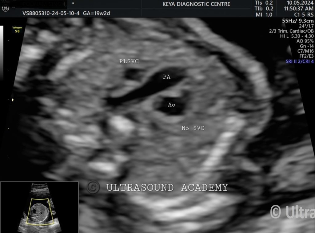
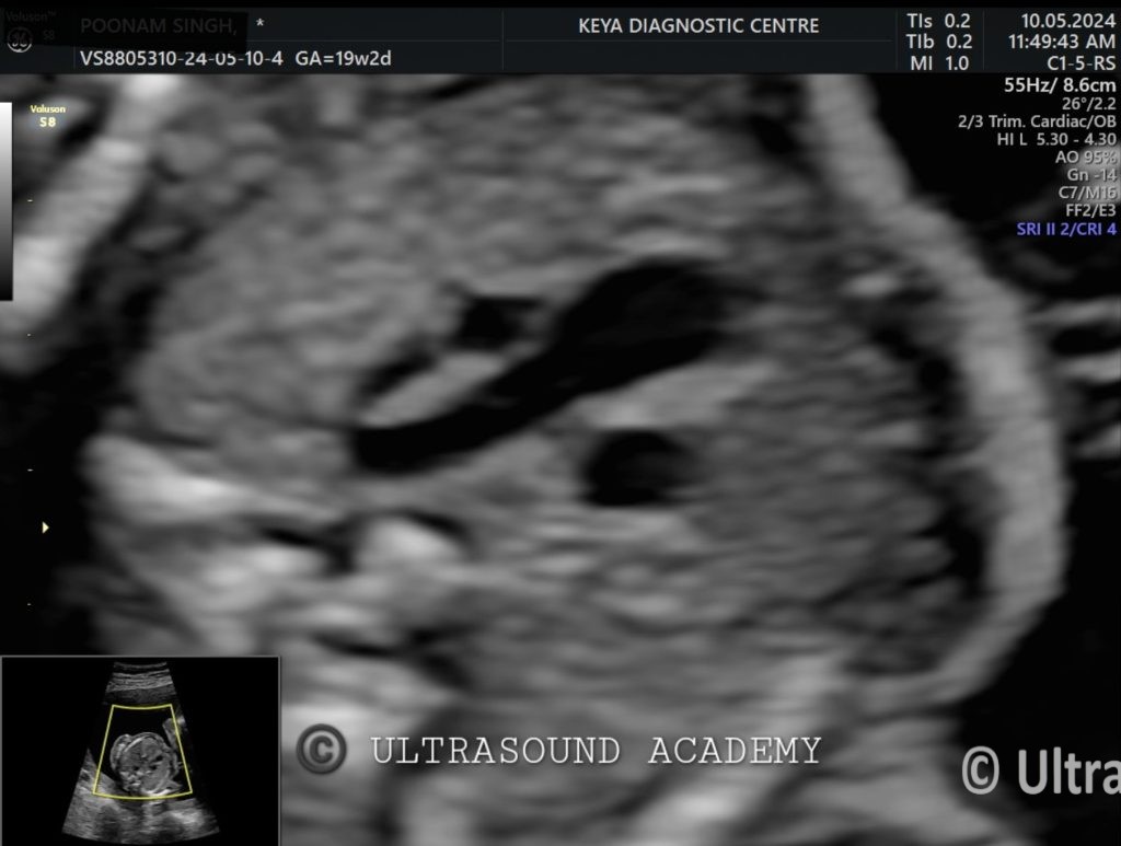
vessel trachea view (3VTV): V sign documented on B mode , representing pulmonary
artery and aorta; But the SVC is seen on left of PA instead of right. Right SVC
is absent.
Coronary
sinus appears dilated.
On Color
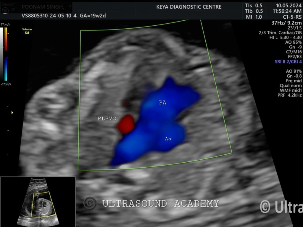
Doppler images and cine: left sided SVC is seen with right brachiocephalic vein.
So,
the diagnosis of Absent right SVC with PLSVC is given.