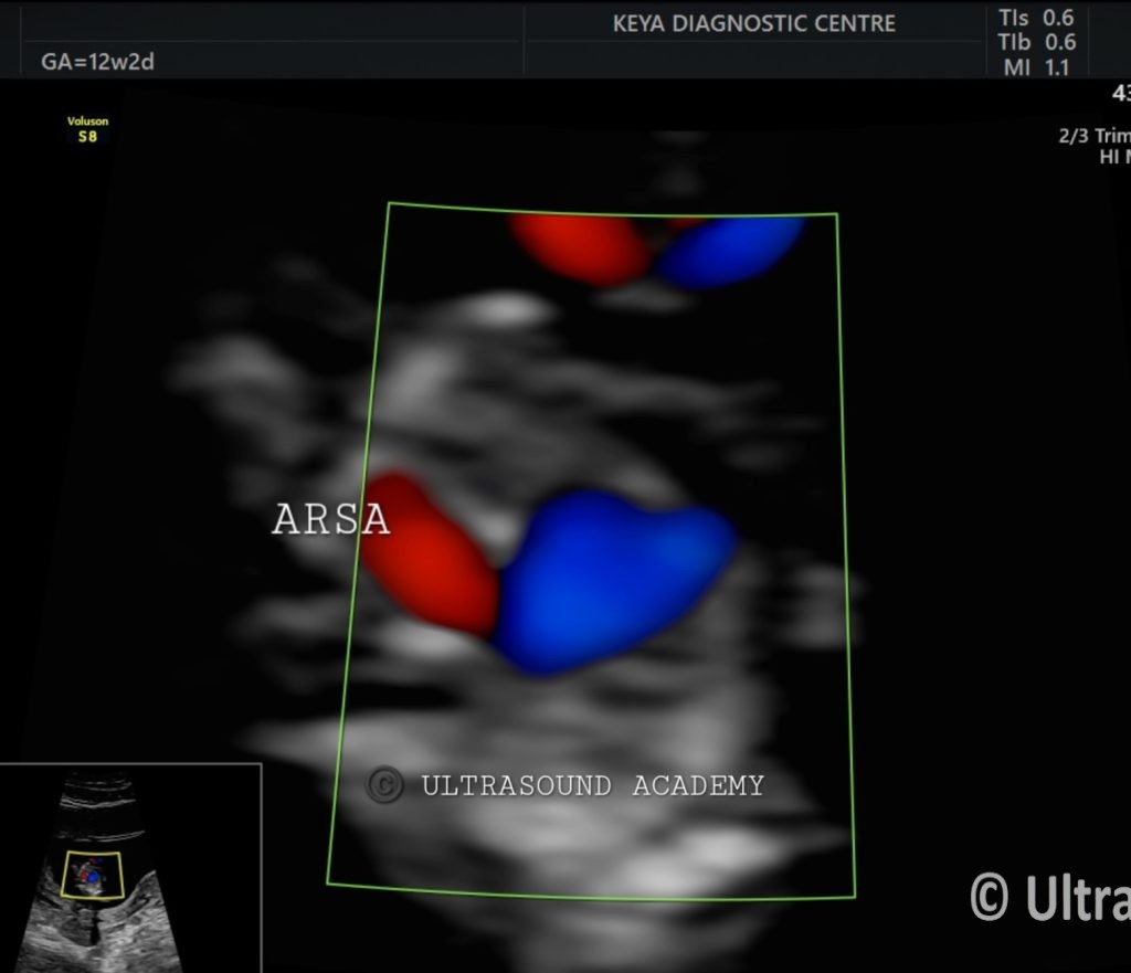
A 30 year-old pregnant female presents for her first trimester ultrasound scan at 12 weeks gestation. She has no significant medical or family history.
Ultrasound Findings:
●The heart appears structurally normal, with a visible four-chamber view. An aberrant right subclavian artery (ARSA) is identified arising from the aorta, noted in the three-vessel trachea view.
●The ARSA is seen arising from the aortic arch and traveling posterior to the trachea and esophagus. This anatomical variation is visualized clearly.
●No other congenital anomalies are seen.
Diagnosis: Isolated aberrant right subclavian artery (ARSA).
Differential Diagnosis:
1. Chromosomal Abnormalities (e.g., Trisomy 21): While ARSA can be associated with chromosomal abnormalities, the absence of other findings significantly reduces the likelihood of this diagnosis.
2. Vascular Ring (Unlikely): ARSA can be associated with a vascular ring causing tracheal or esophageal compression, but this is usually detected postnatally and no evidence of compression is noted in this case.
Discussion:
●The presence of an aberrant right subclavian artery (ARSA) in the first trimester is a known vascular variant where the right subclavian artery arises from the aortic arch, traveling posterior to the trachea and esophagus. ARSA is typically asymptomatic and considered a benign finding when detected in isolation, as in this case.
●Although ARSA can be associated with chromosomal abnormalities such as Down syndrome (Trisomy 21), the risk is substantially lower when no other structural anomalies or soft markers are identified. Therefore, isolated ARSA should be followed up with routine care and serial ultrasounds to monitor fetal growth, without the need for further intervention unless additional concerns arise.