NORMAL CORPUS CALLOSUM AT 23 WEEKS – Case By Dr. Nitin Jadhav
Findings:
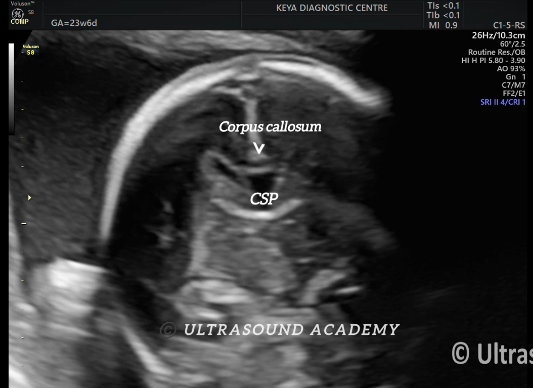
2D as well as 2D demonstrations of corpus callosum at different weeks of gestation are provided here.
Discussion:
· The prosencephalon, during 4–10 weeks of gestation, develops by the process of ventral induction which includes formation, cleavage and midline development. Failure of formation results in aprosencephaly or atelencephaly. Total or partial failure of cleavage results in the spectrum of holoprosencephaly. Abnormal midline development results in agenesis of CC or septal agenesis.
· The corpus callosum (CC) is the largest commissure connecting the two cerebral hemispheres.
· It is a broad plate made up of tightly packed axonal fibres crossing from side to side. In the midsagittal section, the corpus callosum extends from the frontal region anteriorly to overlie the tectum or quadrigeminal plate posteriorly.
· The segments of the CC anterior to posterior are the rostrum, genu, body and splenium. The CC begins to develop at 12 weeks in the region of the genu and progresses posteriorly, forming the body and splenium.
· The rostrum is the last segment to develop.
· The corpus callosal development is complete by 18–20 weeks gestational age.
Key-points:
· Hypoplasia of CC refers to a CC which is normal in length but thin.
· Dysgenesis refers to a CC which is thinner or thicker than normal or partially developed.
· Signs of CC complete agenesis on USG:
1. Absent CSP (cavum septum pellucidum)
2. Teardrop-shaped lateral ventricle
3. The interhemispheric fissure (IHF) is wide giving the “three line” sign
4. The anterior horns are crescent or comma shaped with an outward convexity (“steerhorn” or “Viking helmet” sign)
5. The cingulate sulcus and gyrus are absent
6. The presence of radiating medial hemispheric sulci and gyri is typically seen in the third trimester (“sunburst” sign)
7. Abnormal course of the pericallosal artery is seen on color Doppler
· Signs of CC partial agenesis on USG:
1. Short CSP
2. The length-to-width ratio is termed the CSP ratio. In a short and wide CSP (as in PACC), the ratio is < 1.5.
3. Colpocephaly and teardrop shape of the lateral ventricle
4. Length of the CC is lesser than the fifth percentile or −2SD
5. Cingulate sulcus seen exactly till the CC ends beyond which a few radial sulci may be seen
error: Content is protected !!