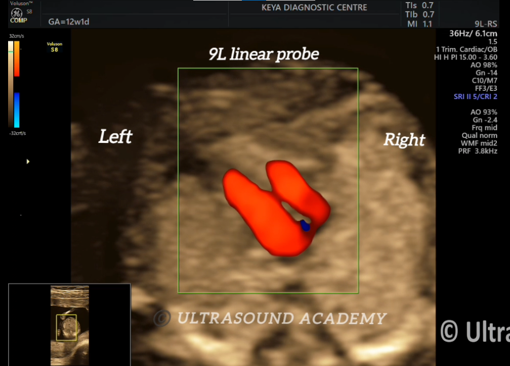
Normal evaluation of fetal heart at 12 weeks antenatal scan is evaluated using a 9L linear transducer which give better resolution than a curvilinear transducer. But, its use is limited due to poor penetration.
Four chamber heart, outflow tracts (RVOT, LVOT), inter-atrioventricular septum, atrio-ventricular valves can be seen on colour doppler.
Discussion:
Normal views to be seen at NT scan are:
4 chamber view
3 vessel view
3 vessel trachea view
LVOT view
RVOT view
Key-points:
On colour doppler any signs of regurgitation, stenosis or valvular dysplasia can be ruled out.
Any regurgitant flow on colour doppler will be seen as aliasing or mixing of colours.
On 3 vessel and 3 VT views, PLSVC or supracardiac TAPVCs can be diagnosed at NT scan.
Ductus venosus must be studied with fetal heart during NT scan.