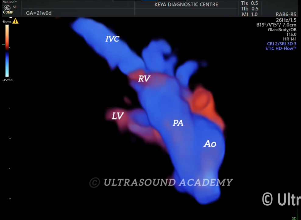
Glass body imaging (volume rendering) cine showing bilateral ventricles giving rise to the outflow tracts.
Discussion:
Glass body imaging, also known as volume rendering or transparent rendering, is an advanced ultrasound technique used in fetal echocardiography to provide a comprehensive view of the fetal heart during an anomaly scan. Let me break down the key aspects of this imaging method:
Technique Overview:
Glass body imaging creates a three-dimensional (3D) visualization of the fetal heart
It allows for a transparent, see-through view of cardiac structures
Helps in detecting subtle structural abnormalities that might be missed in traditional 2D ultrasound
Key Benefits:
Provides a more comprehensive view of cardiac anatomy
Enables detailed examination of spatial relationships between heart structures
Improves detection of complex congenital heart defects
Allows rotation and manipulation of the 3D image for thorough assessment
Typical Timing:
Usually performed during the mid-pregnancy anomaly scan
Typically conducted between 18-22 weeks of gestation