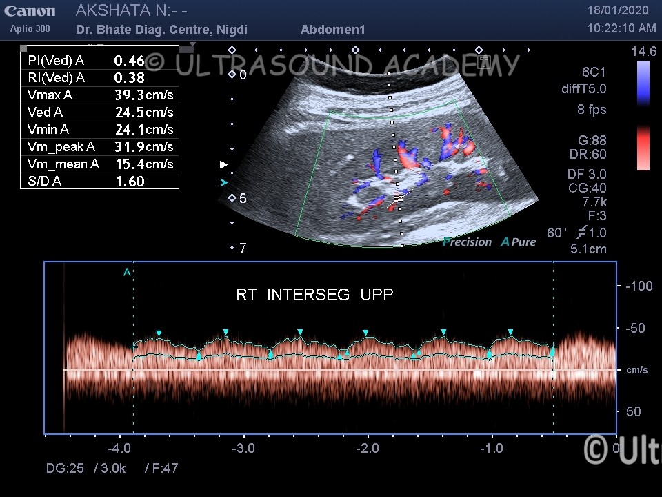
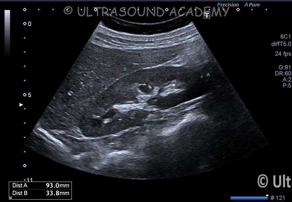
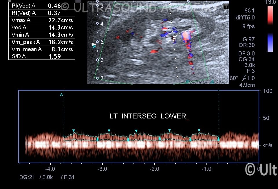
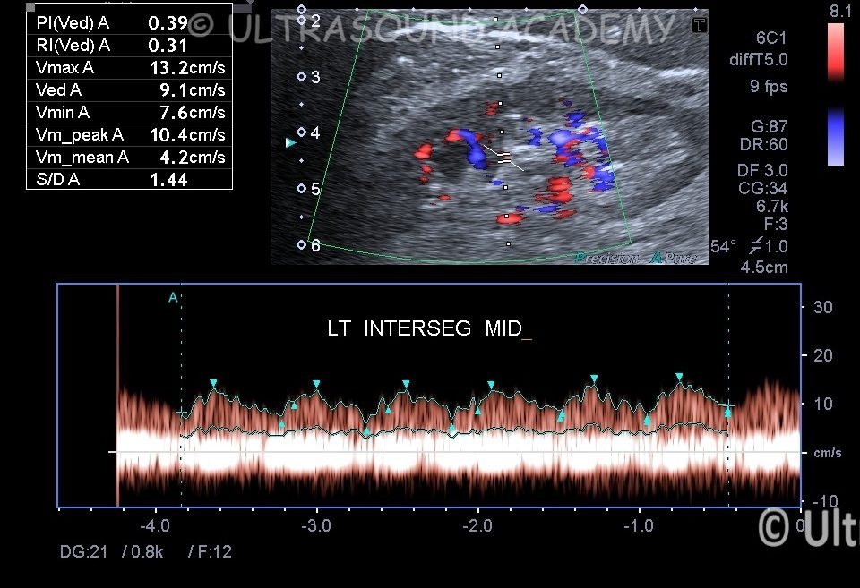
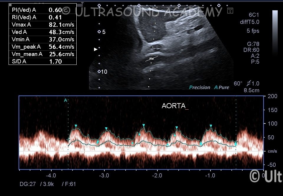
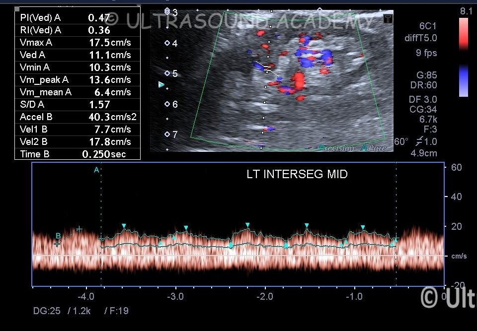
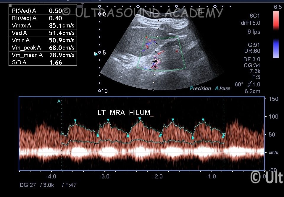
This 25 yr old young female had C/O uncontrolled hypertension.
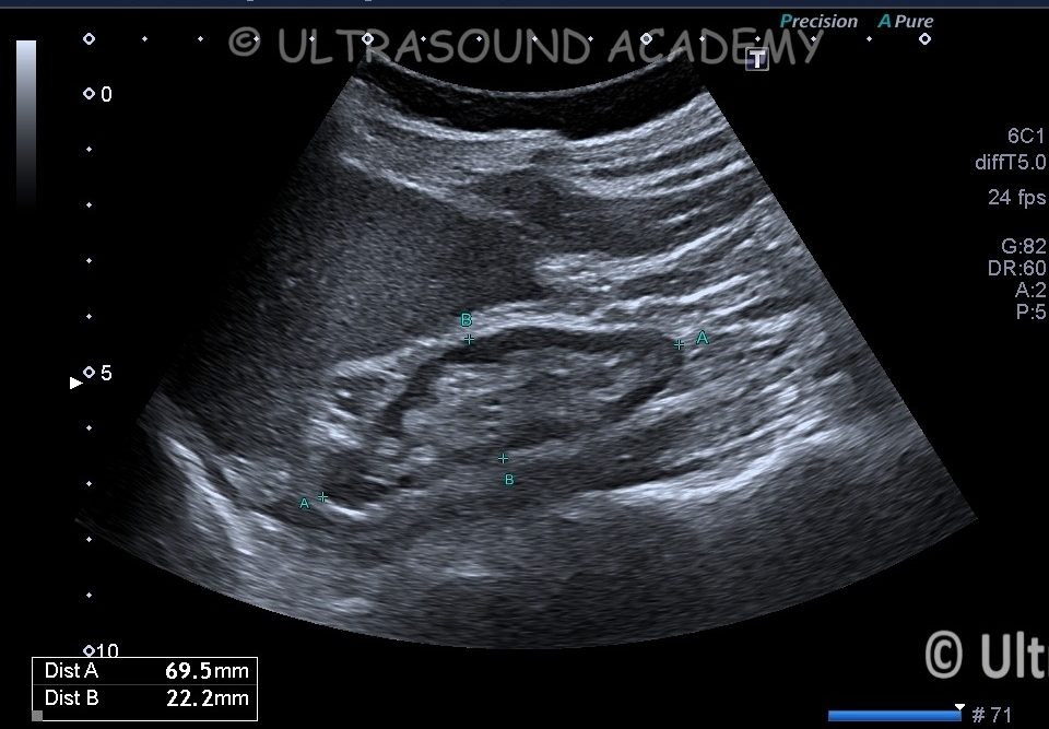
Screening USG abdomen was S/O small left kidney hence renal Doppler was advised.
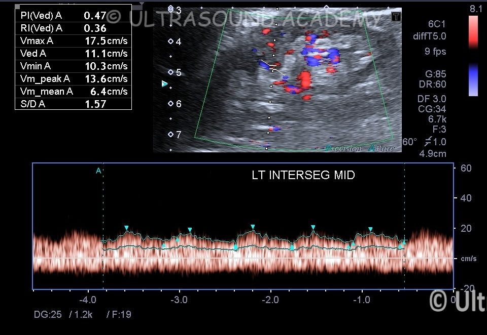
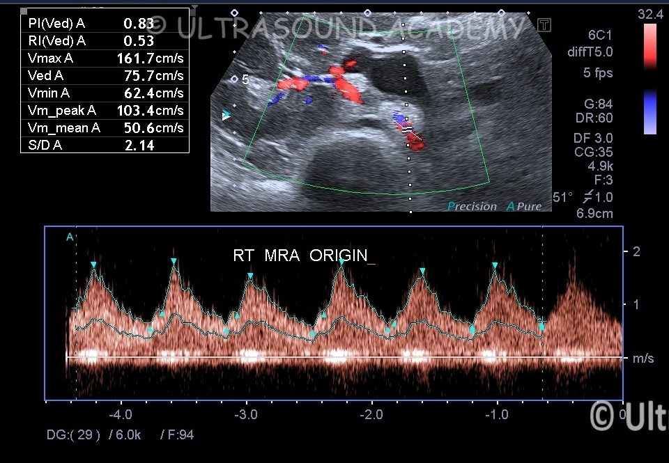
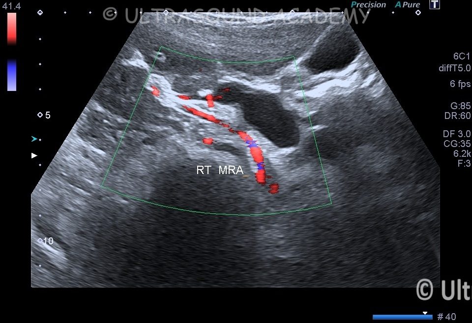
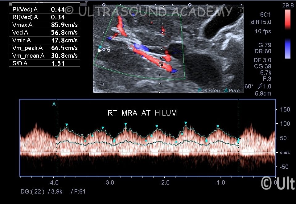
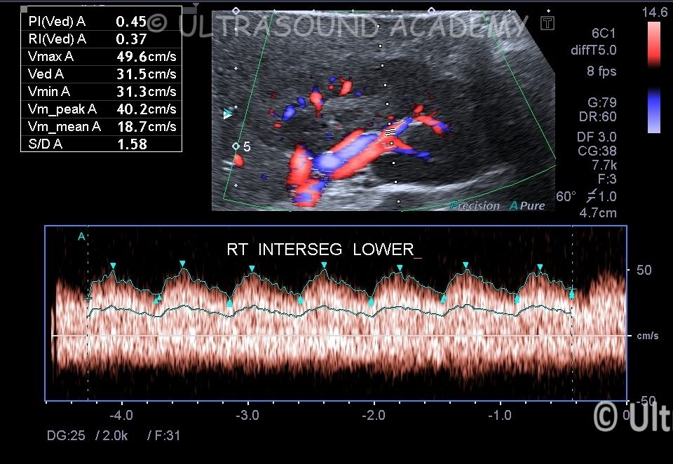
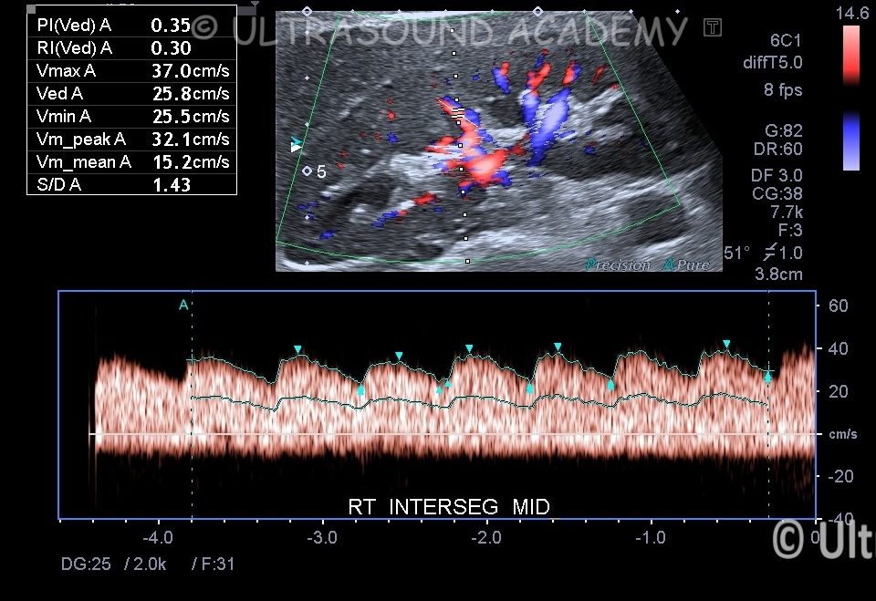
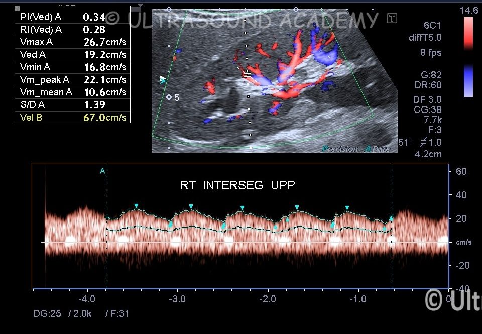
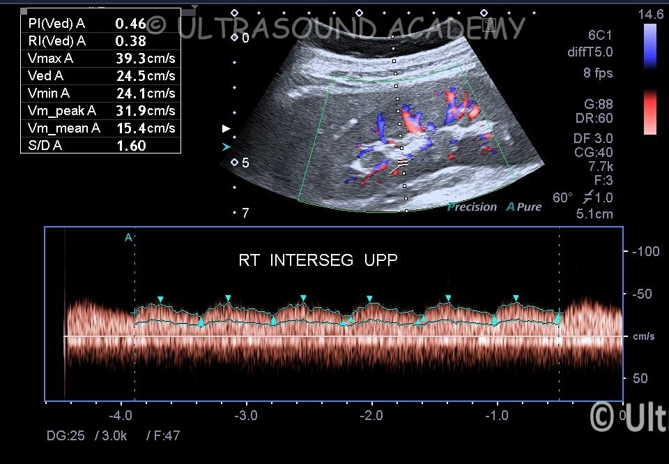
Renal Doppler examination showing low velocity flow in intrarenal as well as main renal arteries in both sides.
All segmental and main Renal arteries shows increased accelerated time with typical parvus tardus pattern waveform.
Even abdominal aorta is also showing low velocity parvus tardus waveform.
On grey scale images visualized part of abdominal aorta was normal raising suspicion for thoracic aorta/ Aortic arch pathology.
CT aortogram was done and showed. Severe narrowing involving thoracic aorta likely due to aortoarteritis.
Key Doppler Findings in Aortoarteritis
Increased Velocity: Due to stenosis or narrowing of arteries, Doppler may show a higher-than-normal velocity of blood flow proximal to the stenotic site.
Turbulent Flow: When stenosis occurs, blood flow becomes turbulent, which can be detected by the Doppler as irregular patterns.
Blunted or Reduced Flow: In more severe cases of aortoarteritis, particularly when there is occlusion, Doppler ultrasound may show little or no flow in the affected arteries.
Flow Disturbance: Inflammation can also lead to irregularities in the smooth laminar flow seen in healthy arteries
Key Findings on Renal Doppler in Aortoarteritis
Renal Artery Stenosis (RAS):
Inflammation of the renal arteries, due to aortoarteritis, can lead to narrowingof the renal arteries, which reduces blood flow to the kidneys. This results in an increase in velocity in the narrowed segment.
Doppler ultrasound can detect the velocity of blood flow through the renal arteries. A peak systolic velocity (PSV) of >180 cm/sin the renal artery is often used as a threshold to suggest significant stenosis.
Post-stenotic turbulence: Beyond the site of stenosis Doppler may reveal turbulent flow, which manifests as chaotic blood flow patterns.
Renal Artery Occlusion:
In severe cases of aortoarteritis, if the stenosis progresses to total occlusion, the Doppler may show absent flowin the renal artery, indicating a lack of blood supply to the kidney.
Renal Impairment:
Renal artery resistive index (RI):A high resistive index (>0.80) in the renal arteries can suggest reduced renal perfusion and possible renal damage.
Renal Artery Aneurysms:
Rarely, aneurysmsof the renal artery may develop in the setting of aortoarteritis. Doppler ultrasound can be used to detect abnormal flow patterns suggestive of an aneurysm.
Doppler Waveform Characteristics in Renal Artery Stenosis
Pre-stenosis flow: The Doppler waveform in the proximal renal artery (before the stenosis) is typically smooth and laminar, with a characteristic high diastolic flow.
Stenosis: When stenosis is present, the Doppler waveform at the site of the narrowing will show increased peak systolic velocities, and the waveform may be systolic sharp with a low diastolic flow.
Post-stenosis flow: Distal to the stenosis, the flow will often appear disturbed or turbulent with a broad, low-velocity diastolic phase.