ACUTE UTERINE PERFORATION – Case By Dr. Nitin Jadhav
Our patient is a 30-year-old woman without any known disease. Recently she underwent dilatation & curettage (D & C) procedure for RPOC. She was referred to us for USG pelvis by the gynaecologist I/V/O severe pelvic pain.
Findings and discussion:
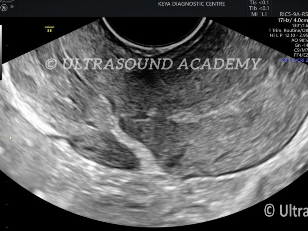
On TVS , uterus is retroverted with normal size , shape and endometrial echo however, there is hyperechoic tract is noted at the anterior uterine wall in the lower segment.
The hyperechoic tract is seen communicating with the peritoneal cavity.
There is no evidence of localized collection at present.
The tract is showing mild vascularity on Color Doppler.
On thorough scanning of adnexa:
Both ovaries and adnexa appear unremarkable at present scan without any evidence of localized collection.
So, the diagnosis of Acute uterine perforation is given.
It is mandatory to accurately diagnose uterine rupture as early as possible , as it can cause haemorrhagic shock and maternal mortality.
Uterine perforation represents a serious complication that can occur as a result of any type of intrauterine procedure.
This patient treated conservatively. Follow up scans were normal.
Iatrogenic uterine perforation refers to an unintentional injury to the uterine wall, often occurring during gynecological procedures such as dilatation and curettage (D&C), hysteroscopy, or intrauterine device (IUD) insertion. Ultrasound (USG) plays a crucial role in diagnosing and managing such cases.
Ultrasound in Uterine Perforation Diagnosis
- Transabdominal Ultrasound (TAUS):
-
- Useful for detecting significant free fluid in the abdominal cavity (e.g., blood or bowel content).
- May show uterine wall defects if they are large.
- Transvaginal Ultrasound (TVUS):
-
- Provides a detailed view of the uterus and surrounding structures.
- Detects the site and extent of the uterine wall defect.
- Identifies displaced intrauterine devices or surgical instruments, if present.
- Reveals any abnormal collection of fluid or blood in the pelvis.
- Key Findings on Ultrasound:
-
- Uterine wall discontinuity or thinning.
- Displaced surgical material, such as a curette, into the myometrium or beyond.
- Free intraperitoneal fluid or hematoma in the pelvis.
- Bowel loops or other structures abnormally close to the uterine wall (indicating perforation into the abdominal cavity).
When to Use Ultrasound
- Suspected uterine perforation based on clinical symptoms such as severe pain, heavy bleeding, hypotension, or signs of peritonitis after a gynecological procedure.
- If there’s concern about abnormal placement of an IUD or other intrauterine devices.
error: Content is protected !!