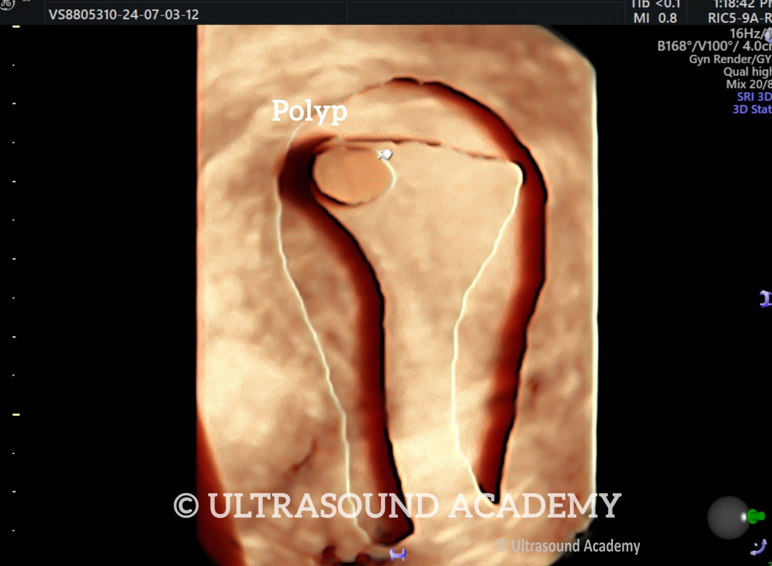
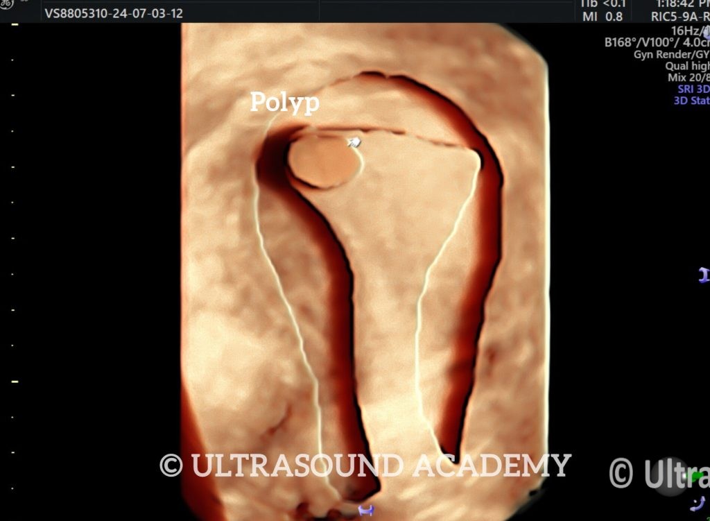
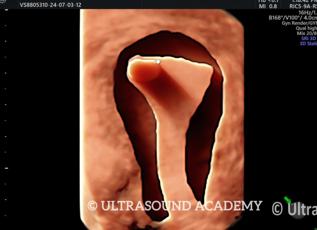
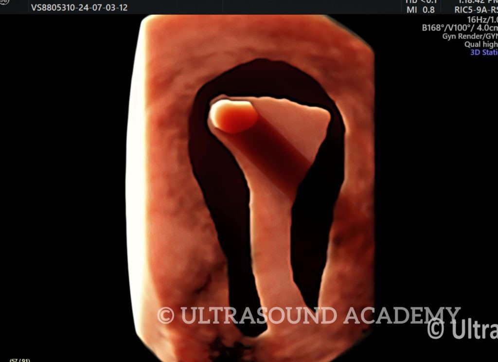
Well defined hyperechoic lesion is seen within endometrial cavity.
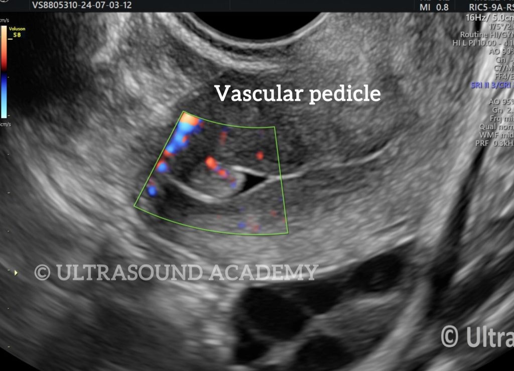
Lesion shows vascular pedicle on Doppler S/O polyp
An endometrial polyp is a localized overgrowth of the endometrial tissue, often detected during a pelvic ultrasound (USG). Here’s what to expect in an ultrasound report for an endometrial polyp:
Ultrasound Features of Endometrial Polyp:
Hyperechoic or Isoechoic Mass: The polyp appears as a well-defined, homogenous, echogenic (bright) mass within the endometrial cavity.
Endometrial Thickening: Focal or diffuse thickening of the endometrium may be seen.
Smooth Contours: Polyps usually have smooth, regular margins.
No Significant Posterior Shadowing: Unlike fibroids, polyps do not typically show acoustic shadowing.
Presence of a Feeding Vessel (Doppler USG): Color Doppler often shows a single central feeding vessel (a distinguishing feature from other lesions like fibroids).
Size and Number: Can range from a few millimeters to several centimeters and may be single or multiple.
Best Ultrasound Techniques for Diagnosis:
Transvaginal Ultrasound (TVS): Offers a clearer and more detailed view of the endometrium.
Saline Infusion Sonohysterography (SIS): Involves injecting sterile saline into the uterus to improve visualization of the polyp and differentiate it from other causes of endometrial thickening.
Differential Diagnosis:
Endometrial hyperplasia (diffuse thickening, no focal mass).
Submucosal fibroids (hypoechoic with shadowing, may distort the endometrium).
Endometrial carcinoma (irregular mass with vascularity).
Management:
Small, asymptomatic polyps may be monitored.
Symptomatic polyps (causing abnormal bleeding or infertility) are removed via hysteroscopic polypectomy.
Biopsy is recommended for postmenopausal women or if malignancy is suspected.