60 years old postmenopausal female referred by gynaecologist with c/o PV spotting
Findings and discussion:
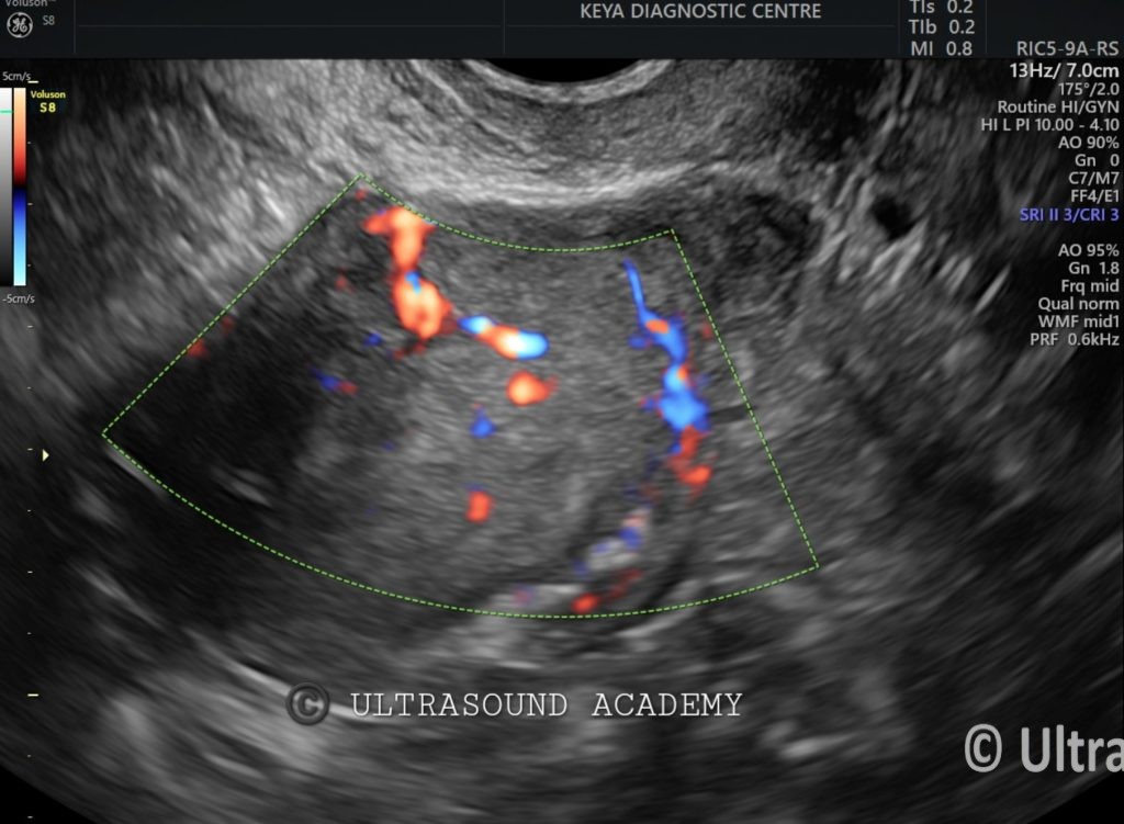
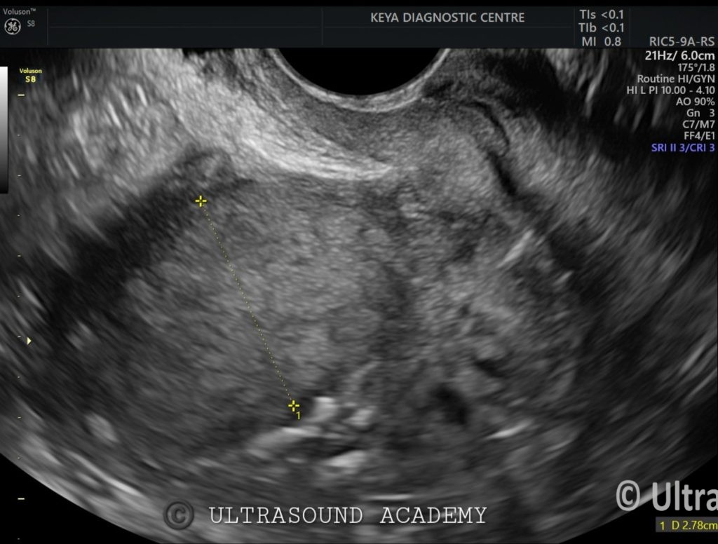
On B mode ultrasound : endometrial echo is grossly thickened measuring approximately
27-28 mm.
On Color Doppler : significantly high vascularity seen within the thickened endometrium.
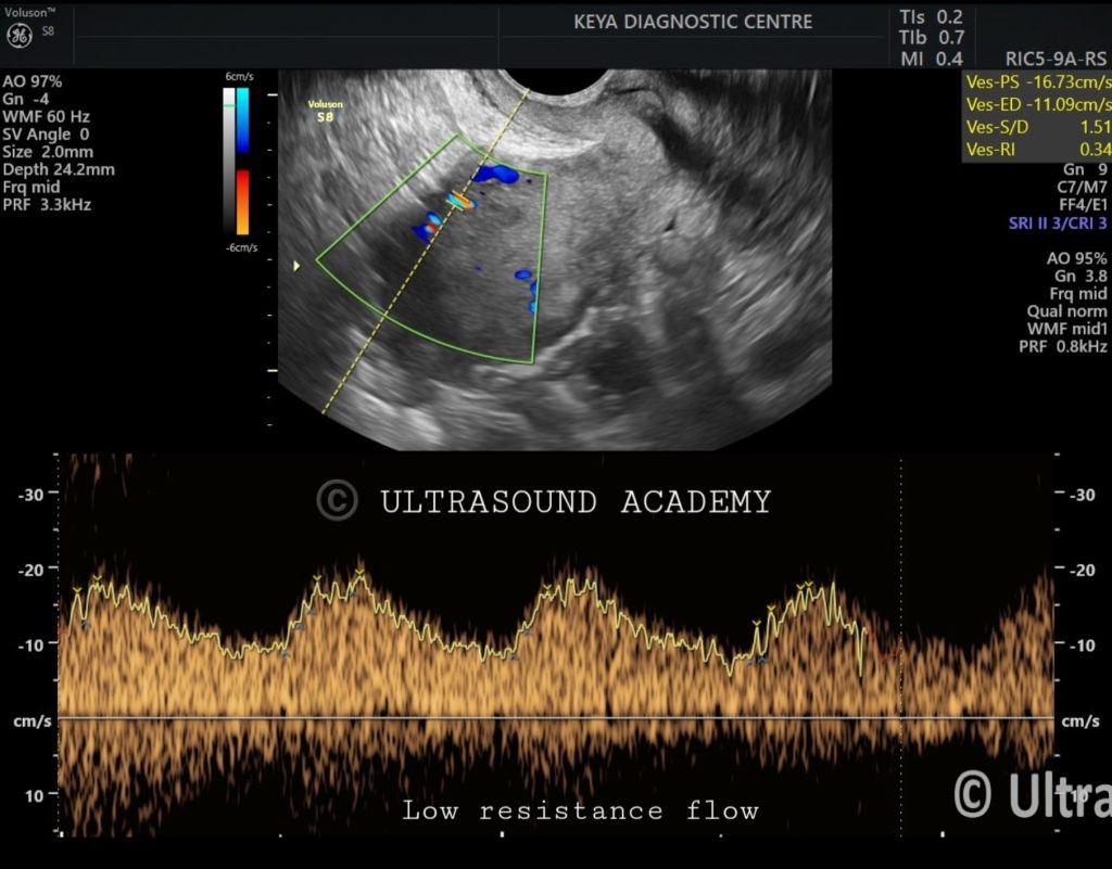
On pulsed wave (PW) Doppler : endometrial vessels show low resistance waveform with RI value of 0.38.
There is no obvious myometrial thinning or extension beyond myometrium is appreciated.
These findings favors neoplastic etiology.
Ca endometrium confirmed on postoperative histopathology.
So, the take home points for Ca endometrium:
I) Postmenopausal female with thickened endometrium
II) significantly high vascularity
III) Low resistance waveform on PW doppler.
Ultrasound is one of the primary imaging tools used to evaluate endometrial carcinoma . It helps assess the thickness, vascularity, extension beyond myometrium
Ultrasound:
Transabdominal Ultrasound (TAS):
Provides an overview of the uterus and adnexa
Transvaginal Ultrasound (TVS):
More detailed than TAS for endometrium , myometrium and adnexa
USG Findings Suggestive of Endometrial Carcinoma:
Increased Endometrial Thickness:
Postmenopausal women: >5 mm is concerning.
Premenopausal women: Thickness varies, but abnormal thickening (e.g., >15 mm) may indicate malignancy.
Irregular Endometrial Echotexture:
Heterogeneous or irregular endometrial lining.
Disruption of the normal interface between the endometrium and myometrium.
Fluid in the Endometrial Cavity:
May be due to malignancy especially in postmenopausal women.
Poorly Defined Endometrial-Myoendometrial Junction:
Suggests invasive cancer spreading into the uterine muscle layer.
Presence of Mass or Polypoid Lesion:
A focal lesion or endometrial mass may be seen.
Increased Vascularity on Doppler Imaging:
Higher vascularity the lesion with low resistance waveform raise suspicion of malignancy.
OTHER INVESTIGATIONS :
Endometrial Biopsy or Dilation & Curettage (D&C) for histopathological confirmation.
MRI or CT Scan for staging and to assess local and distant spread.