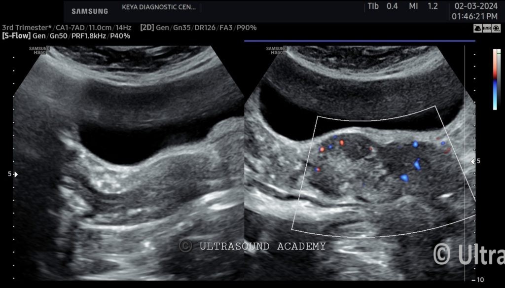
Elderly female presents with postmenopausal vaginal bleeding and pelvic pain. The patient is referred for ultrasound evaluation of the pelvis for suspected cervical malignancy.
Ultrasound Findings and Description:
An enlarged, heterogeneous mass lesion involving both lips of cervix and vaginal walls is seen.
It is hypoechoic with irregular borders.
Increased vascularity noted within the mass on color Doppler imaging, indicative of neovascularization.
The mass appears to invade the parametrium but no definitive bladder or rectal involvement noted.
Normal appearance of the endometrium. Ovaries are not well visualized, consistent with postmenopausal status. No free fluid in the pelvis.
Diagnosis: Carcinoma of the cervix, likely invasive cervical carcinoma.
Differential Diagnosis:
Cervical Polyp: These can present as focal cervical masses but are usually well-defined, without aggressive features or vascularity.
Cervical Fibroid (Leiomyoma): A rare possibility in this location, cervical fibroids are well-defined and more homogeneous compared to the findings in this case.
Conclusions:
Large, hypoechoic cervical mass with irregular borders and hypervasculaity suggestive of cervical carcinoma.
Further evaluation with biopsy and advanced imaging (MRI pelvis) is recommended to assess local invasion and staging.