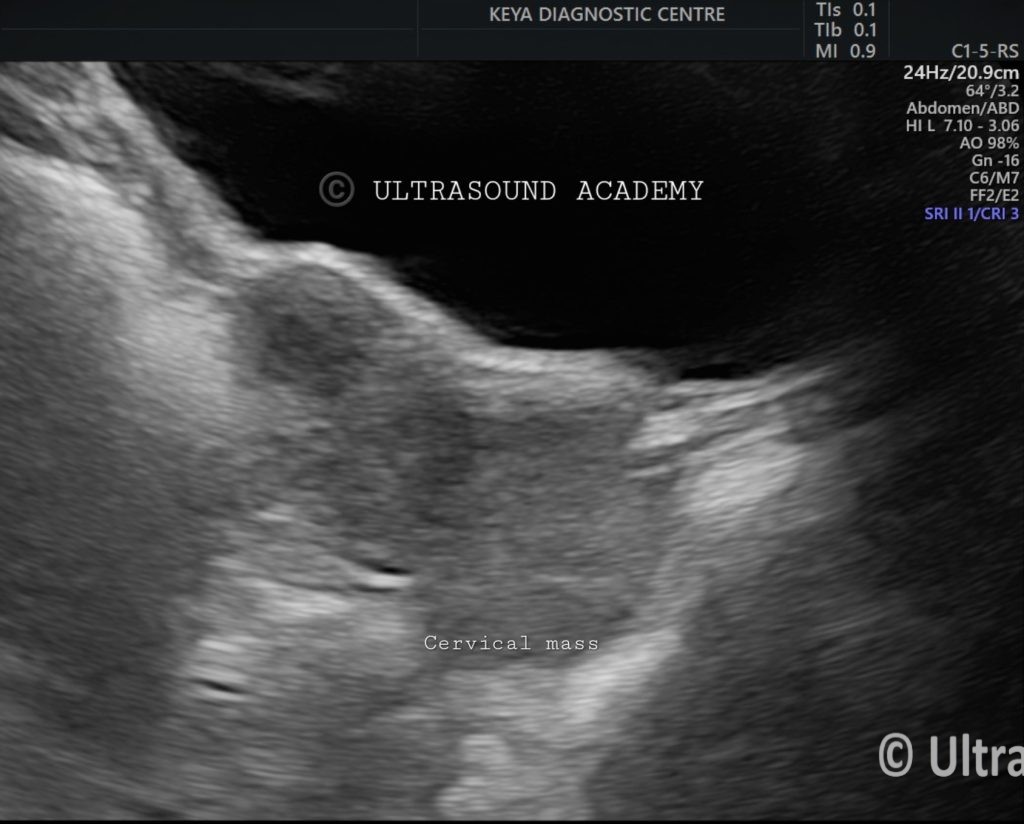
Sagittal and axial transvaginal scan shows lobulated,huge mass along the cervix.
A cervical mass is seen slightly indenting but not involving the urinary bladder
There is an increased internal vascularity.
Conclusion:
The investigation approach for cervical cancer involves a combination of screening tests Pap smear, HPV testing, imaging MRI, CT, PET-CT,Transvaginal,Transabdominal Ultrasound and biopsy(gold standard) to establish the diagnosis, stage the disease, and plan appropriate treatment. MRI is particularly preferred for detailed local staging of cervical cancer, while PET-CT helps in evaluating distant metastasis.
For cervical cancer diagnosis and treatment, ultrasound plays a key role in detecting and assessing the extent of the tumor, especially in the early stages. This modality helps evaluate tumor size, involvement of adjacent organs like the bladder or rectum, and lymph node metastasis, making it a useful tool, especially when MRI is not available.
Ultrasound characteristics in cervical cancer include:
Size of the tumor (< 4 cm or > 4 cm).
Parametrial invasion.
Invasion into the vagina or adjacent organs.
Hydronephrosis, indicating more advanced disease (Stage IIIB).
While MRI is considered the gold standard for detailed staging.In clinical practice, ultrasound is often used alongside other imaging modalities such as MRI and CT for comprehensive staging.
Differential Diagnosis:
Cervical polyp: Can present as a mass but typically non-invasive. Further imaging might show a benign appearance.
Leiomyoma: Fibroid arising from the cervix, possibly causing distortion of the cervical anatomy.
Endometrial cancer: While less common, endometrial carcinoma can cause displacement of structures in advanced stages.