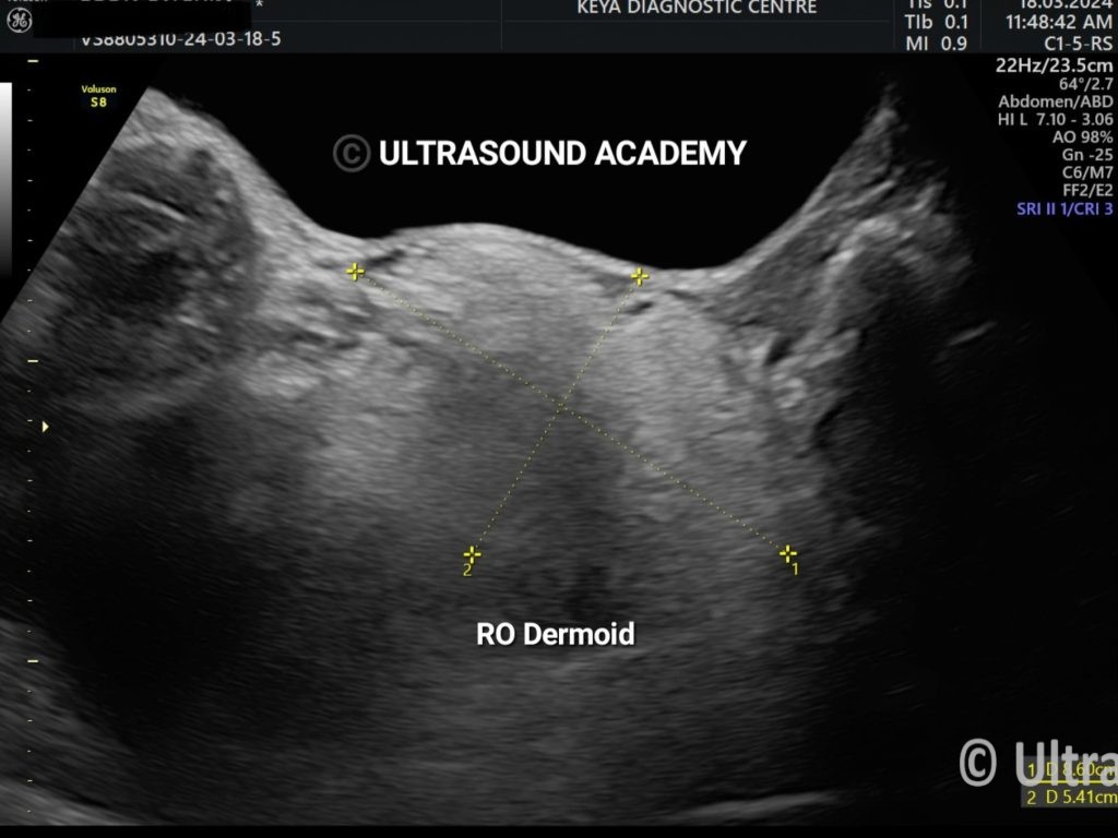
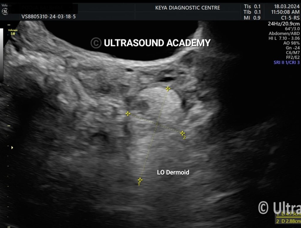
A middle age female, presented with pain in lower abdomen, since last 2-3 months.
Findings:
Two well-defined hypoechoic cystic lesions were seen with densely echogenic nodules within it and with multiple thin, echogenic bands were also noted within. No internal vascularity was evaluated on colour doppler imaging.
The diagnosis of bilateral ovarian dermoid cyst was made on ultrasonography.
Discussion:
Dermoid cysts are ectodermal inclusion cysts containing skin and skin appendages; sebaceous glands, hair follicles, and sweat glands.
A Rokitansky nodule or dermoid plug refers to a solid protuberance projecting from an ovarian cyst in the context of mature cystic teratoma. It often contains calcific, dental, adipose, hair, and/or sebaceous components. This region has the highest propensity to undergo malignant transformation.
Epidermoid cyst, also ectodermal in origin, are lined solely by squamous epithelium. Dermoids, instead, contain tissues derived by at least two of the embryonic layers.
Key-points:
·Well-defined cystic ovarian lesion with solid mural components within.
·Echogenic thin bands (dot and dash sign).
·Dermoid plug (echogenic content with post acoustic shadowing).