28 years old female referred by gynaecologist for ultrasound pelvis in view of infertility.
Findings and discussion:
On B mode TVS ultrasound and cine:
Uterus is retroverted and is normal in size.
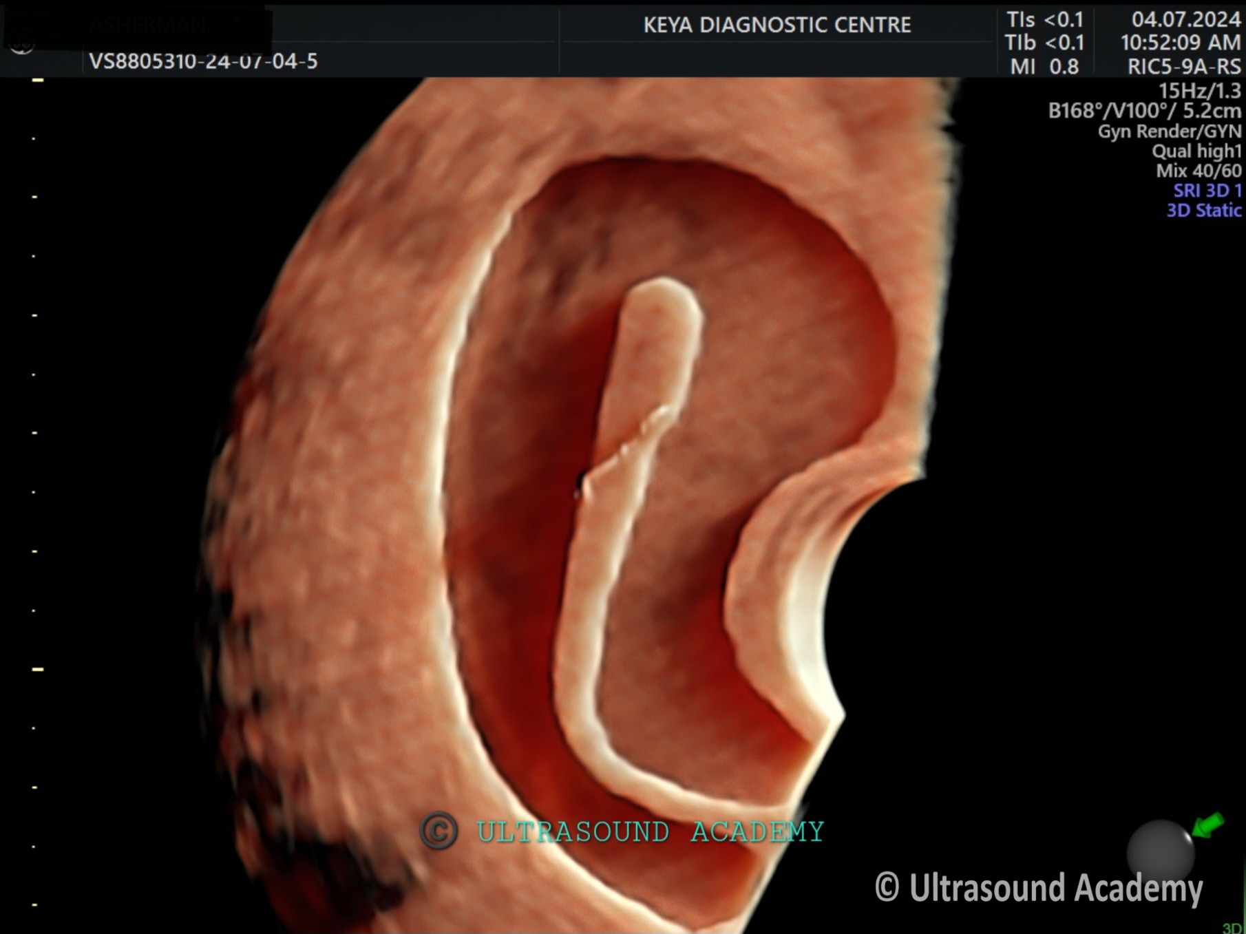
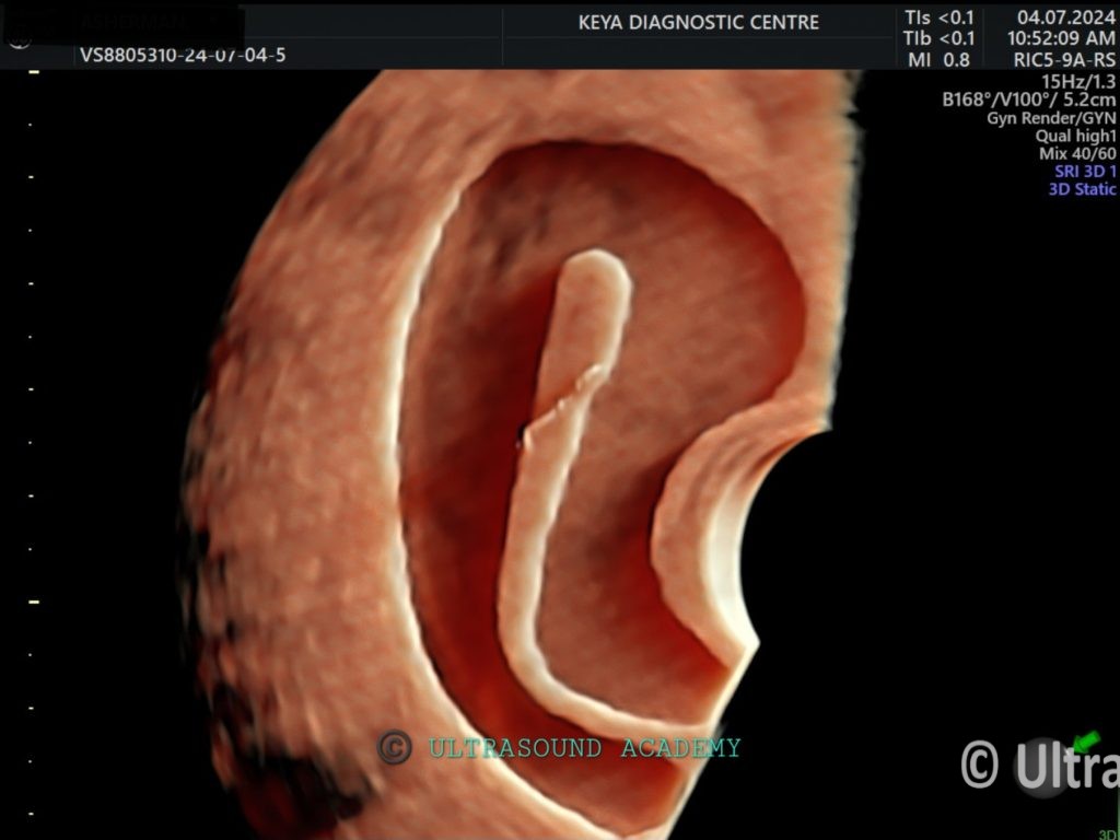
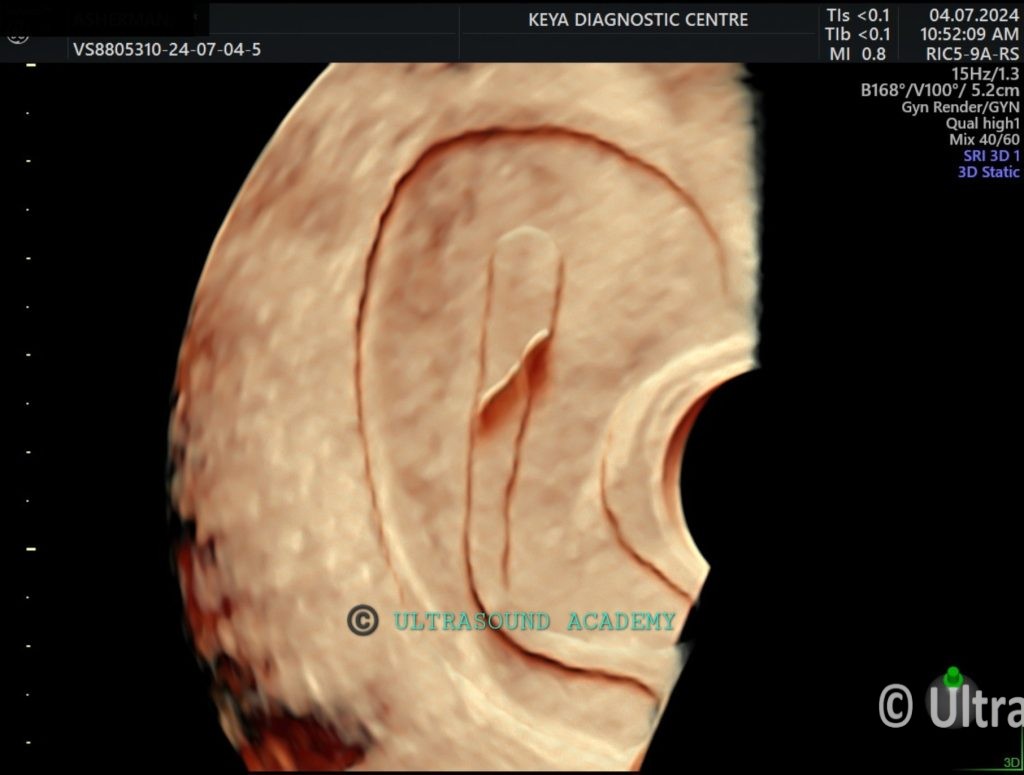
There is hypoechoic synechiae band is seen in midline in the endometrial canal on the sagittal and axial section.
Synechiae band seems to dissect or bifurcate the endometrial cavity.
Synechiae band shows vascularity on Color Doppler.
Both the ovaries are seen and appear normal.
No evidence of hydrosalpinx seen.
On 3D/4D transvaginal ultrasound:
Interrupted endometrial line is very well appreciated.
So, the diagnosis of uterine synechiae band is given which can be one of the cause of infertility in this patient.
Asherman syndrome is a condition characterized by intrauterine adhesions (scar tissue) that can lead to menstrual irregularities, infertility, or recurrent miscarriages. Ultrasound (USG) findings can help in the diagnosis, especially when combined with other imaging techniques like saline infusion sonohysterography (SIS) or hysterosonography.
Ultrasound (USG) Findings in Asherman Syndrome
Thin or Irregular Endometrium
The endometrial lining appears abnormally thin or irregular, especially in the mid to late proliferative phase when it should normally thicken.
Intrauterine Adhesions (Synechiae)
Bright echogenic bands or fibrous strands within the endometrial cavity, sometimes seen as non-mobile structures.
These bands may not allow the endometrial lining to expand properly.
Distorted or Obliterated Uterine Cavity
The normal endometrial cavity shape may appear irregular or partially/completely obliterated due to adhesions.
Poor Endometrial Response to Hormonal Stimulation
If the patient undergoes estrogen therapy, the endometrial thickness does not increase as expected.
Saline Infusion Sonohysterography (SIS) Findings
SIS improves visualization of adhesions as non-filling areas where saline fails to distend the cavity.
The normal endometrial cavity should distend symmetrically with fluid, but in Asherman syndrome, adhesions prevent full expansion.
Doppler Ultrasound Findings
In some cases, color Doppler may show reduced vascularity in the affected areas, indicating fibrotic tissue.
Other Diagnostic Methods
Hysteroscopy (Gold Standard): Direct visualization of intrauterine adhesions.
MRI: Can provide additional details on the extent and nature of adhesions.
Hysterosalpingography (HSG): Shows filling defects in the uterine cavity.
If Asherman syndrome is suspected on ultrasound, further evaluation with hysteroscopy or SIS is often recommended for confirmation and treatment planning.