Neonate referred by paediatrician with complains of large head size.
Referred for Neurosonogram:
Findings and discussion:
On B mode ultrasound:
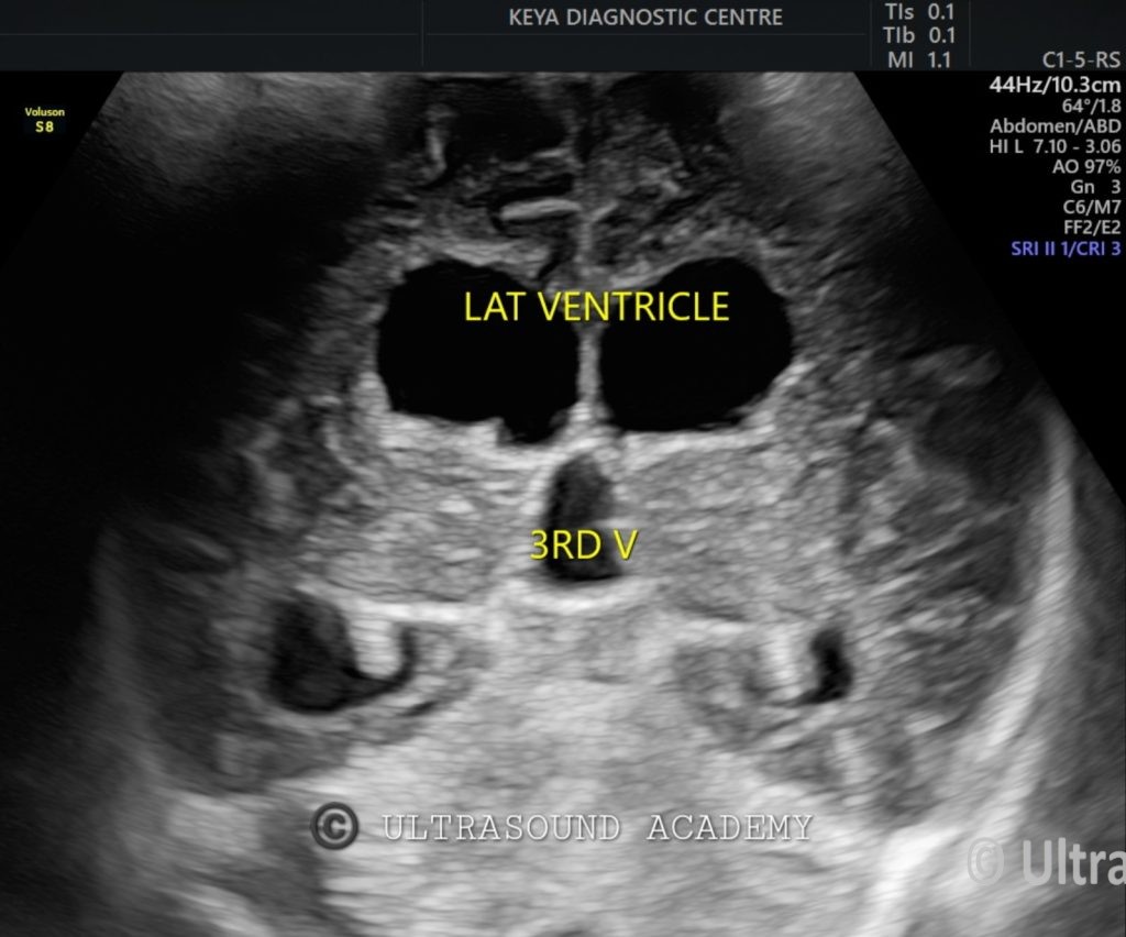
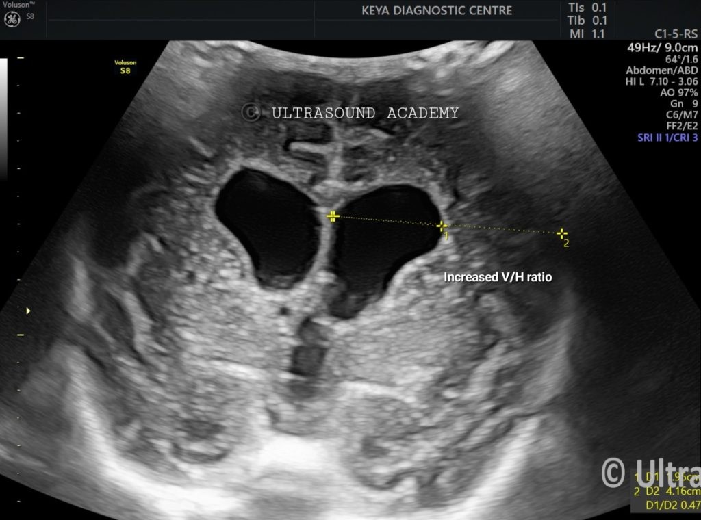
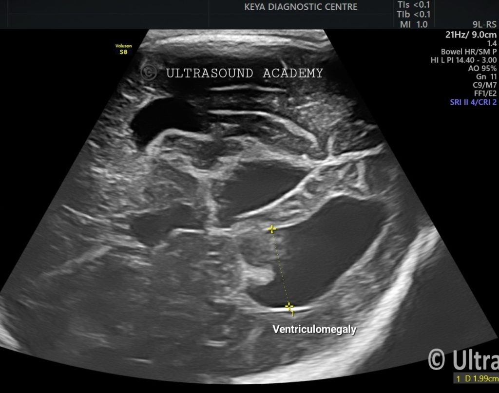
Both the lateral ventricles and third ventricle is dilated.
Diameter of lateral ventricle is measured at parieto-occipital fissure with maximum diameter reaching up-to 20 mm s/o ventriculomegaly.
Also, there is increase in the V/H ratio measuring 0.40 (normal range: 0.20 to 0.25)
Fourth ventricle is normal in size.
Mega cisterna magna is noted.
Thinned out corpus callosum is seen.
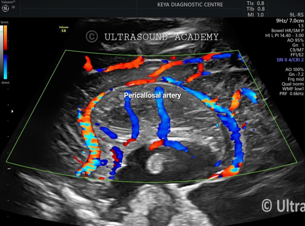
On Color Doppler: Pericallosal artery is well visualized.
So, in view of dilatation of bilateral lateral & third ventricle with normal fourth ventricle and thinned out corpus callosum diagnosis of Aqueduct stenosis is given.
Aqueductal Stenosis in Neonates: Ultrasound Findings and Diagnosis
Aqueductal stenosis is a condition where the cerebral aqueduct (Sylvian aqueduct) is narrowed or blocked, leading to obstructive (non-communicating) hydrocephalus in neonates. Ultrasound (USG), particularly cranial ultrasound through the anterior fontanelle, is a key imaging modality for early detection.
Ultrasound Findings in Neonatal Aqueductal Stenosis
Ventriculomegaly (Hydrocephalus):
Dilated lateral ventricles (frontal and occipital horns).
Dilated third ventricle (key finding).
Normal or collapsed fourth ventricle (since the blockage is above it).
Thinning of Brain Parenchyma (due to chronic hydrocephalus in severe cases).
Dangling Choroid Plexus:
Floating in the enlarged ventricles due to CSF accumulation.
Increased Resistive Index (RI) in Doppler Studies:
Elevated RI (>0.8) in the pericallosal or anterior cerebral arteries suggests increased intracranial pressure.
Diagnosis and Further Imaging
Cranial USG is the first-line imaging for neonates.
MRI Brain (preferred for confirmation) to evaluate aqueductal narrowing, possible causes (congenital vs. acquired), and periventricular changes.
Causes of Aqueductal Stenosis
Congenital: X-linked hydrocephalus (L1CAM mutation), Chiari II malformation, Dandy-Walker malformation.
Acquired: Post-hemorrhagic or post-inflammatory causes.