A 16 year-old female presents with intermittent pelvic pain and dysmenorrhea. She has no history of prior surgeries or known uterine anomalies. Referred for ultrasound evaluation due to persistent pelvic discomfort and abnormal menstrual patterns.
Ultrasound Findings :
The uterus is normal in size and shape. Both uterine cornua are well visualized.
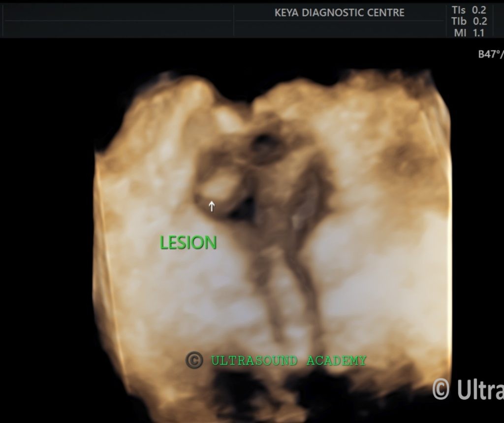
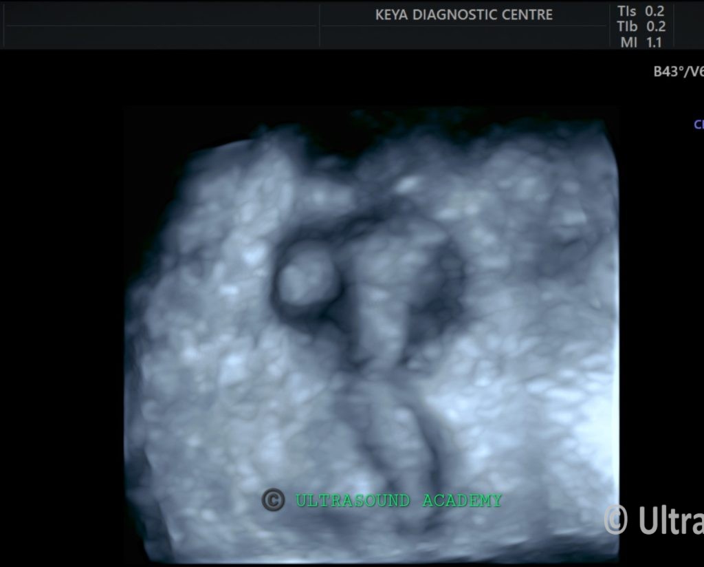
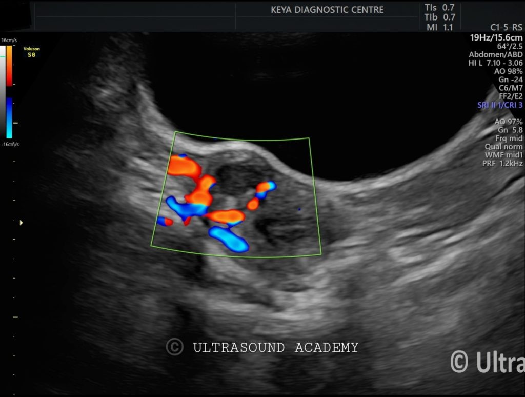
A well-defined, intramural isoechoic lesion is seen near the fundus of the uterus, close to the right cornu. The lesion measures approximately 2 cm in diameter.
The lesion contains a central cystic area with fine internal echoes, suggestive of blood products or debris.
Predominantly peripheral vascularity is noted on Doppler imaging. No communication with the endometrial cavity is identified.
The endometrial lining is intact and normal in thickness with no invasion or distortion by the lesion.
Both ovaries are normal in size and appearance.
No free fluid seen in the pelvis.
Diagnosis: Accessory and Cavitary Uterine Mass (ACUM).
Differential Diagnosis:
Juvenile Cystic Adenomyoma: Similar imaging findings with central cystic areas typically presents in younger patients with painful menstruation.
Intramural Fibroid with Cystic Degeneration: Although typically more hypoechoic, intramural fibroids can undergo cystic degeneration, producing similar findings.
Endometriotic Cyst (Endometrioma): Endometriotic cysts may present with cystic lesions containing echoes due to blood products, but they are usually located in the ovaries rather than within the uterus.
Discussion:
The ultrasound findings of a well-defined intramural lesion with a central cystic component, peripheral vascularity, and lack of communication with the endometrial cavity are most consistent with an Accessory and Cavitary Uterine Mass (ACUM).
ACUM is a rare Müllerian anomaly characterized by the presence of a cavitated uterine mass, commonly found near the cornu and unconnected to the main endometrial cavity.
Juvenile cystic adenomyoma is another important differential diagnosis, typically occurring in younger women and presenting with a similar lesion involving the myometrium. The presence of blood products in the central cystic area could suggest menstrual blood accumulation within the cavity. The absence of communication with the endometrial cavity helps differentiate these conditions from other uterine anomalies or fibroids.
Further imaging, such as MRI, may provide better characterization of the lesion and help confirm the diagnosis. Treatment may involve surgical excision, particularly if the mass is causing significant pain or menstrual abnormalities.