ACCESSORY HORN 3D – Case By Dr. Nitin Jadhav
History:
A 29-year-old female presents for routine follow-up of an early pregnancy. She reports no significant symptoms and is asymptomatic. Previous imaging revealed a suspected uterine anomaly. She is referred for ultrasound to assess fetal growth.
Ultrasound Findings:
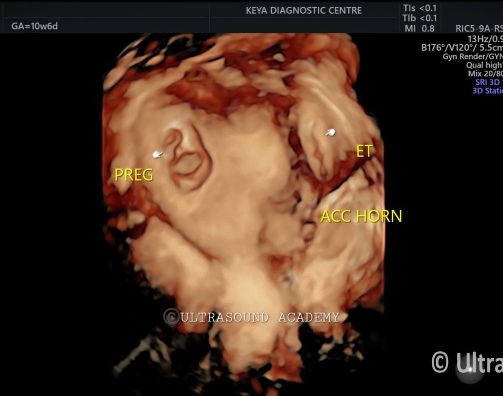
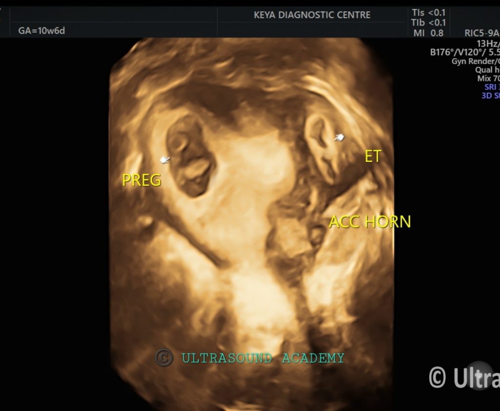
● Uterus (2D and 3D Imaging): Bicornuate uterus visualized. A normal intrauterine pregnancy (IUP) is present in the right horn, corresponding to 10 weeks 6 days of gestation. The crown-rump length (CRL) measures 38 mm.
● 3D ultrasound provides clear visualization of the bicornuate uterus, confirming the two distinct uterine horns.
● A non-communicating left rudimentary horn contains a pseudo sac, measuring 2.7 cm, without a fetal pole or yolk sac.
● The left ovary is normal, measuring 3.2 cm, and is visualized separately from the rudimentary horn with no signs of torsion.
● The sliding sign is positive, indicating the accessory rudimentary horn moves independently of the left ovary.
● No free fluid is seen in the pelvis.
Diagnosis:
Normal intrauterine pregnancy (10 weeks 6 days) in the right horn of a bicornuate uterus, with a pseudo sac in the non-communicating left rudimentary horn.
Differential Diagnosis:
1.Pseudo Sac in Rudimentary Horn: The pseudo sac in the left horn likely represents a fluid-filled structure, often confused with a gestational sac.
2. Hematometra in Rudimentary Horn: The collection could represent retained blood (hematometra), often seen in non-communicating uterine horns.
3.Ectopic Pregnancy (Unlikely): Although unlikely given the absence of a fetal pole, ectopic pregnancy remains a differential diagnosis.
Discussion:
● This case demonstrates a viable intrauterine pregnancy in the right horn of a bicornuate uterus, with a pseudo sac present in the left non-communicating rudimentary horn.
● Bicornuate uterus is a congenital Müllerian duct anomaly, where the uterus is divided into two separate horns.
● The pseudo sac in the left horn is a common finding in such anomalies, often mistaken for an ectopic pregnancy.
● The 3D ultrasound provides detailed visualization of the uterine anomaly, aiding in diagnosis.
● The positive sliding sign confirms that the rudimentary horn moves separately from the left ovary, ruling out ovarian involvement. Regular monitoring is recommended to ensure the normal progression of the pregnancy and to assess any potential complications related to the rudimentary horn.
error: Content is protected !!