21 years old female referred by gynaecologist with c/o cyclical lower abdominal pain / dysmenorrhoea
Findings and discussion:
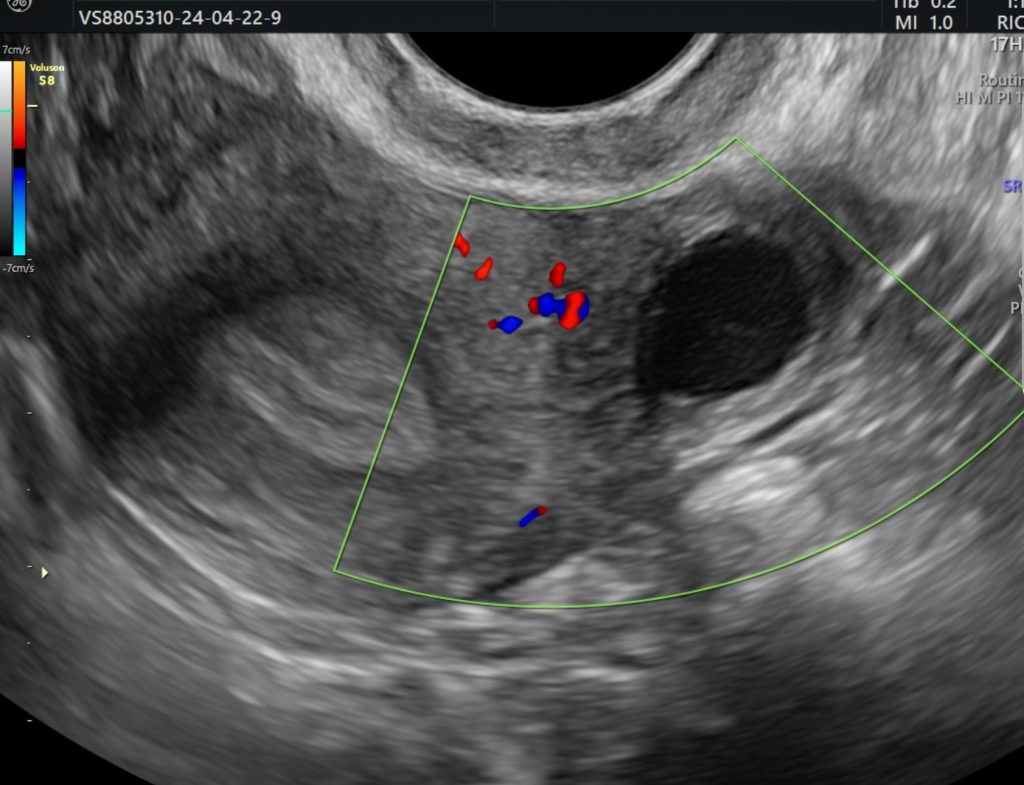
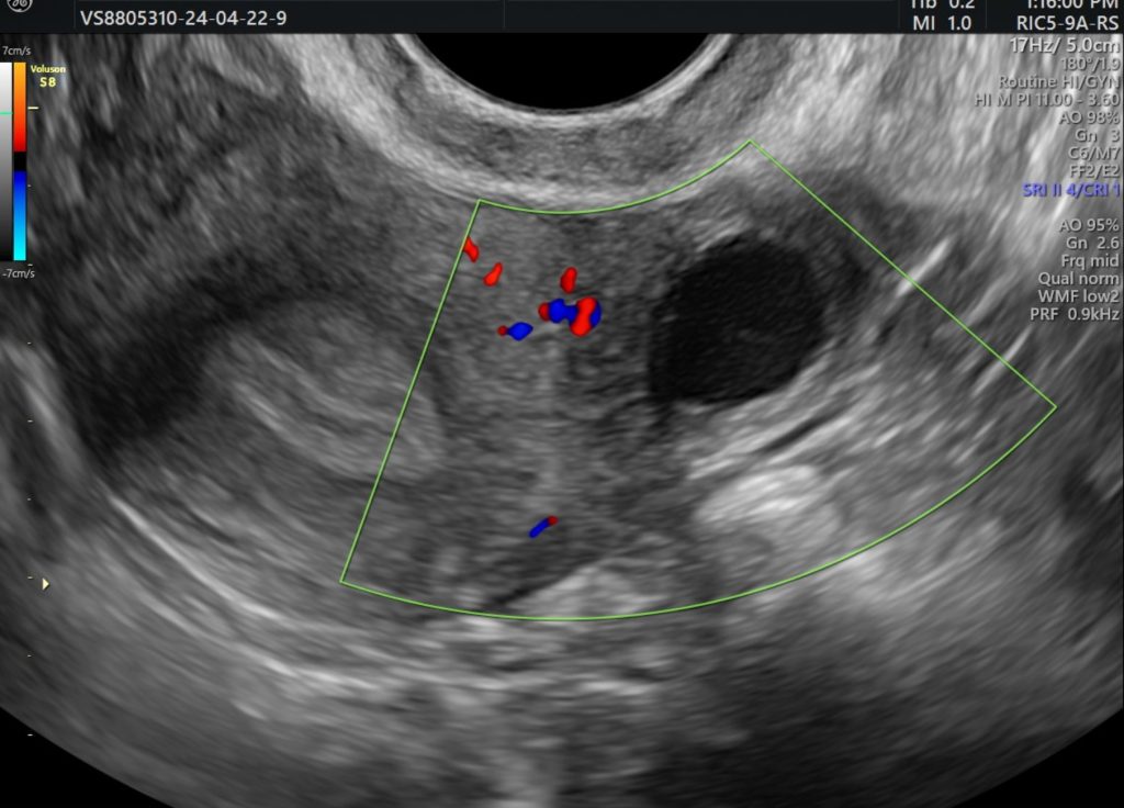
On B mode TVS ultrasound image and cine:
Uterus is anteverted.
Well defined lesion is seen abutting the left cornua of uterus and appears isoechoic to myometrium.
No communication is seen with endometrial cavity.
Lesion shows well defined central cystic area which shows fine echoes within probably representing blood products.
Both the cornua of uterus are well visualized.
Both ovaries are normal and seen separately.
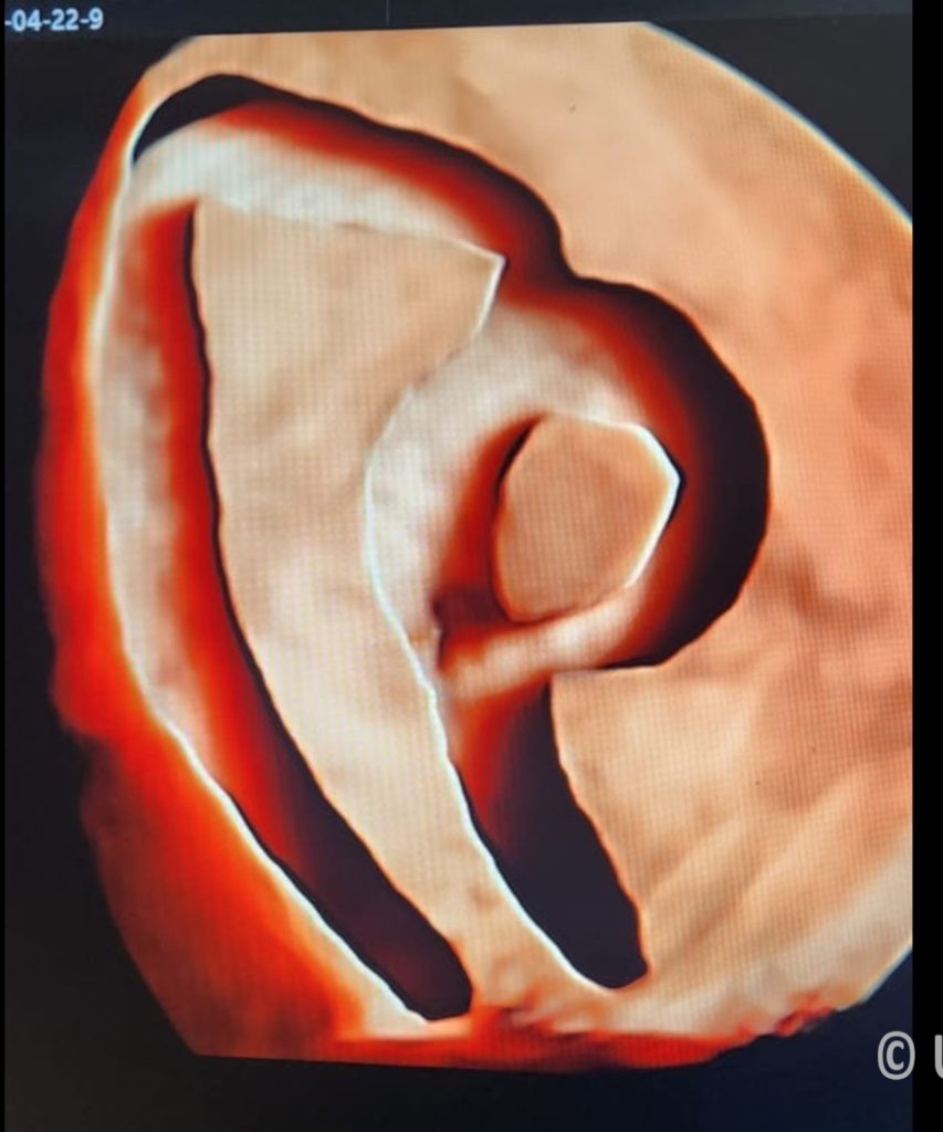
On 3D/ 4D ultrasound this mass is well demonstrated.
So, diagnosis of ACUM (accessory and cavitated uterine mass) , also known as non-communicating accessory uterine cavity.
An Accessory Cavitated Uterine Mass (ACUM) is a rare congenital uterine anomaly that presents as a non-communicating mass within the uterus, often mistaken for other gynecological conditions like fibroids or endometriosis. It is typically associated with severe dysmenorrhea and chronic pelvic pain in young women.
Ultrasound Findings of ACUM
On transabdominal or transvaginal ultrasound, ACUM may appear as:
Hypoechoic or Heterogeneous Mass – A well-defined lesion near the uterine wall, usually at the anterior or lateral part of the uterus.
Cystic Areas – The mass may have cystic spaces filled with hemorrhagic content, which can change over time due to cyclic bleeding.
Non-communicating with the Endometrial Cavity – Unlike a normal uterine cavity or a functional endometrial cyst, ACUM is an isolated structure.
No Myometrial Involvement – It is separate from the endometrium and myometrium, differentiating it from adenomyosis.
Doppler Findings – May show peripheral vascularity but usually lacks central vascularity.
Differential Diagnoses
Fibroids (Leiomyoma) – Usually more solid with posterior acoustic shadowing.
Adenomyosis – Typically diffuse, ill-defined, and involves the myometrium.
Endometrioma – Found in ovaries, not within the uterine wall.
Müllerian Anomalies – Other forms of uterine malformations like a non-communicating horn.
Confirmatory Diagnosis
MRI: Provides better characterization of the lesion, showing hemorrhagic contents and no connection with the endometrial cavity.
Histopathology (after surgical removal): Confirms the presence of endometrial glands, stroma, and smooth muscle.
Management
Medical Treatment: Hormonal therapy (OCPs, progestins, GnRH analogs) may be tried but is often ineffective.
Surgical Excision: Laparoscopic or hysteroscopic resection is the preferred treatment for symptom relief and fertility preservation.