Meckel’s diverticulitis with Sealed off perforation
Findings and discussion-:
This is a 8 yr old child having complaints of right Iliac fossa pain.
This child was having similar complaint 1 month ago for which he underwent appendicectomy.
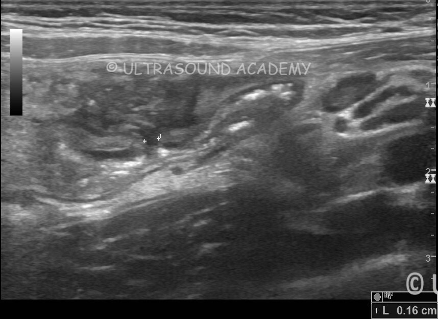
USG abdomen shows long tubular blind ending structure with gut wall signature at RIF. This structure was seen arising from terminal ileum confirming at as Meckel’s diverticulum.
Small discontinuity is seen within the wall of Meckel’s diverticulitis at its base S/O perforation. Small thick collection is seen adjacent to the perforation.
Meckel’s diverticulitis was confirmed on CT
Meckel’s diverticulitis can be challenging to diagnose on ultrasound (USG) due to its often nonspecific findings. However, in suspected cases, USG may reveal the following features:
Typical Ultrasound Findings:
Blind-Ending Pouch:
A tubular or cystic structure located in the right lower abdomen, arising from the ileum.
It does not exhibit peristalsis like normal bowel loops.
Wall Thickening:
Thickened walls of the diverticulum, suggesting inflammation.
May appear hypoechoic due to edema.
Surrounding Inflammatory Changes:
Hyperechoic fat stranding around the diverticulum.
Possible localized fluid collection or abscess formation.
Doppler Study:
Increased vascularity of the diverticular wall on color Doppler (hyperemia), indicative of inflammation.
Intestinal Obstruction Signs:
Dilated bowel loops proximal to the diverticulum in cases where obstruction is associated.
Free Fluid:
Presence of localized or free peritoneal fluid in the pelvis or around the diverticulum in cases of perforation.
Challenges with USG:
Small size and variable position of Meckel’s diverticulum can make it difficult to visualize.
It may be confused with appendicitis, Crohn’s disease, or other ileocecal conditions.
Additional Imaging:
If Meckel’s diverticulitis is suspected but not clearly visualized on USG, CT scan or Meckel’s scan (Technetium-99m pertechnetate) may be more sensitive for confirming the diagnosis.