Findings and Discussion:
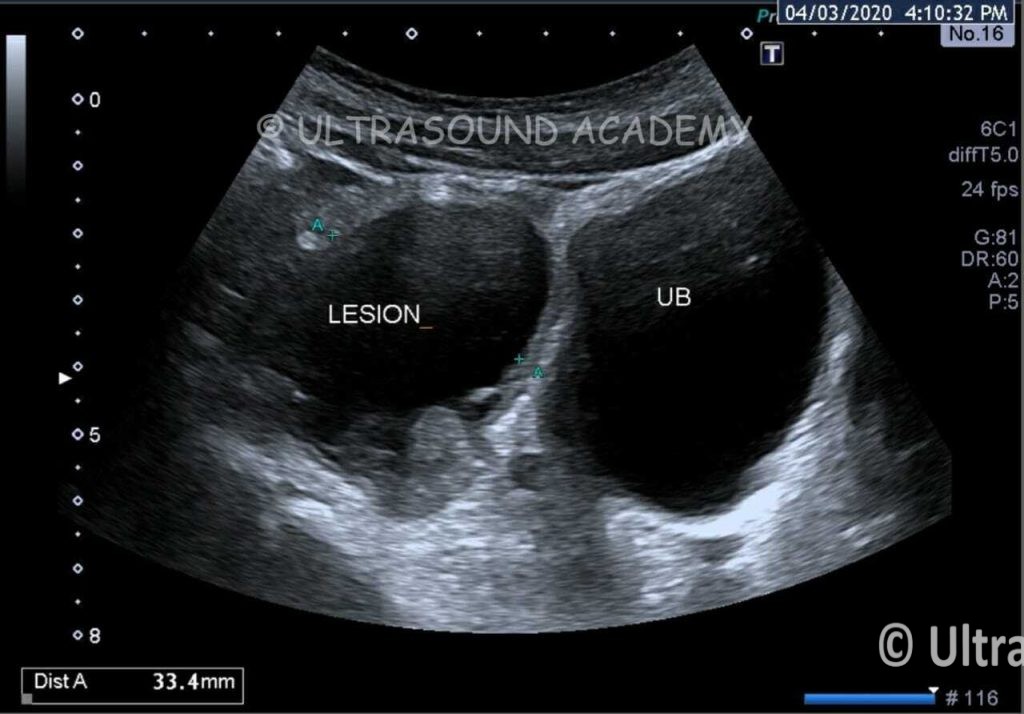
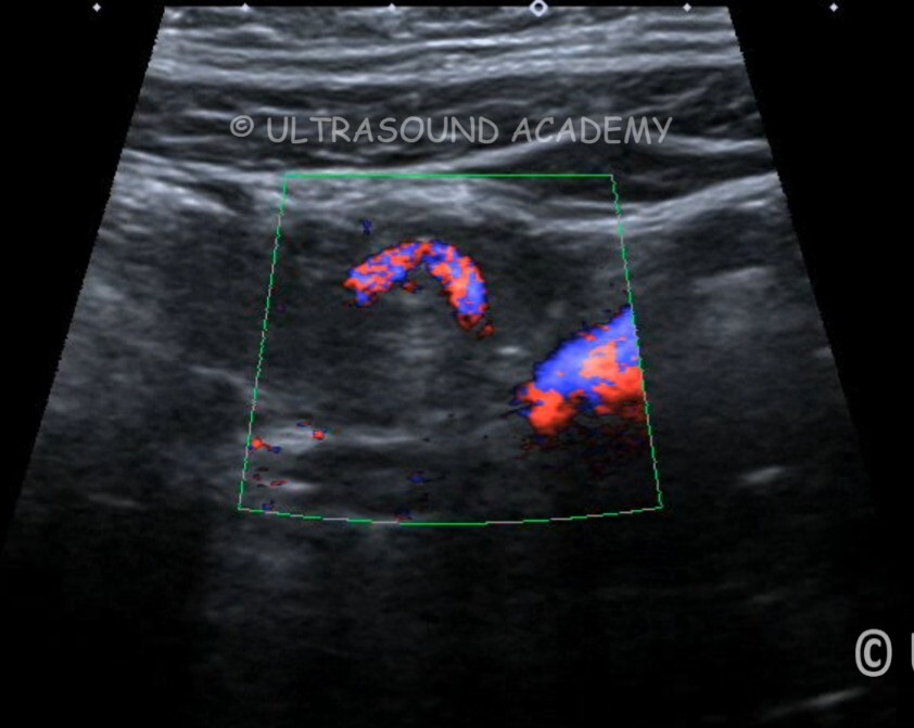
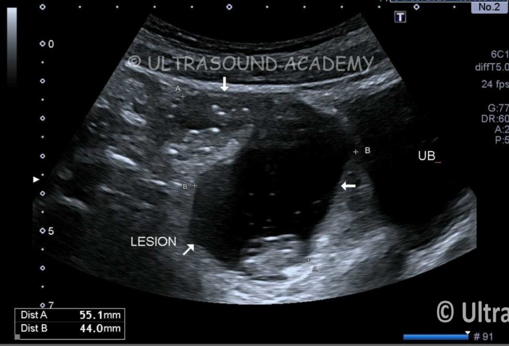
Cystic lesion is seen at RIF containing echogenic debris and air specks.
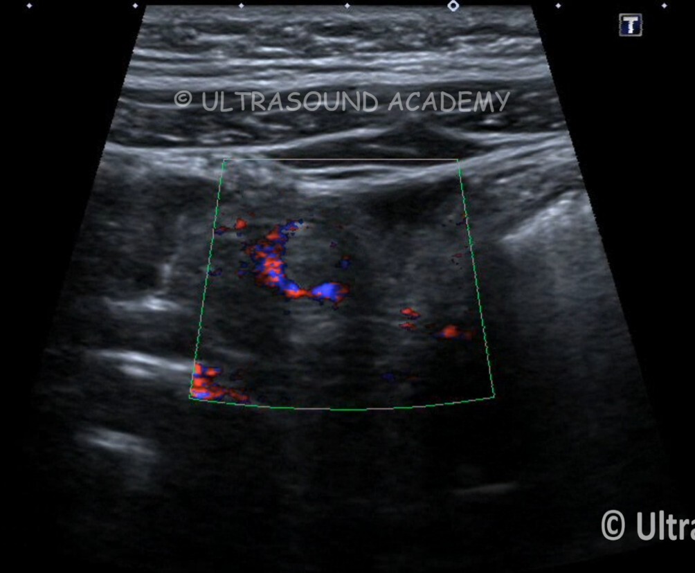
Twisting with whirlpool sign is seen at the pedicle of the lesion.
Lesion is in close proximity with ileal loops.
No obvious continuity with bowel loop could be demonstrated on ultrasound.
Top differentials –
-Meckel’s diverticulitis
-Infected Enteric duplication cyst
-Closed loop obstruction.
-Mucocele of appendix
-localised infected collection etc.
Considering ultrasound findings of gut wall signature, Intraluminal gas, close proximity to terminal ileum and twisting of pedicle, it was reported it as
Torsion of Meckel’s diverticulum with DD of ileal volvulus.
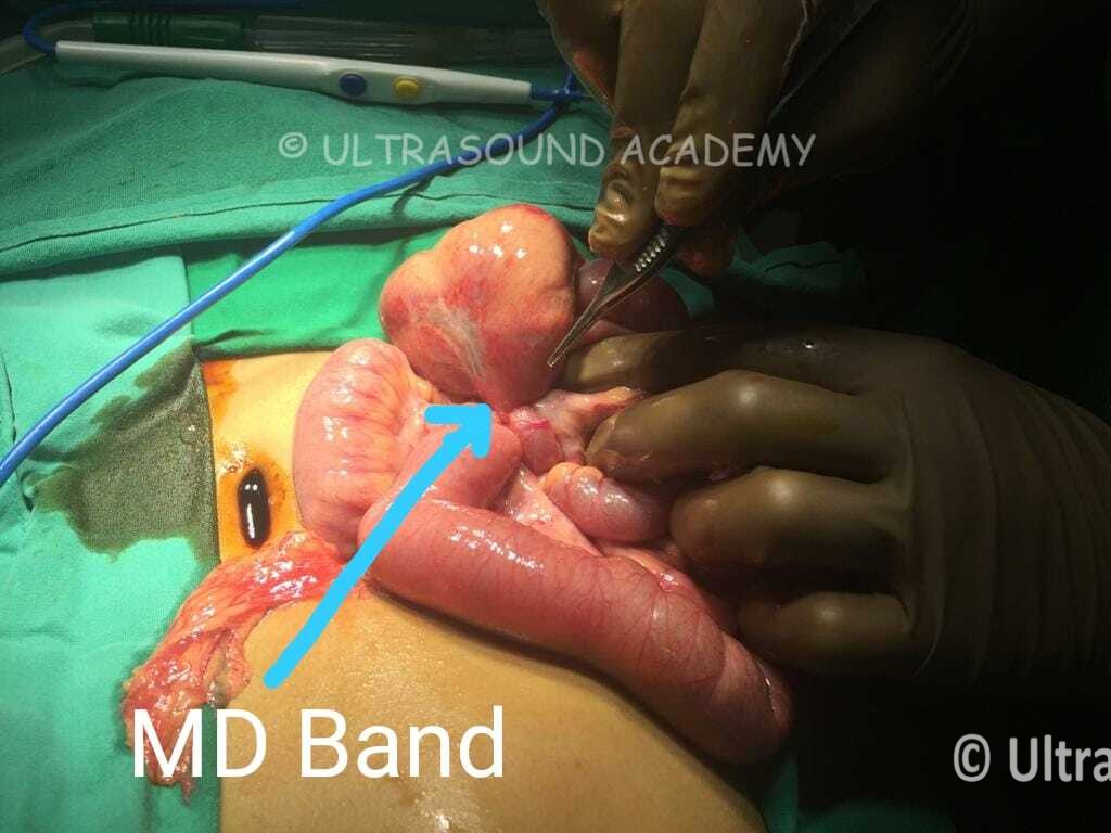
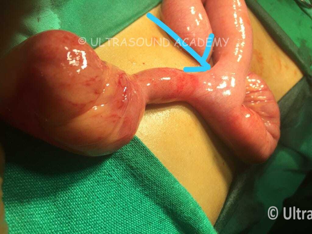
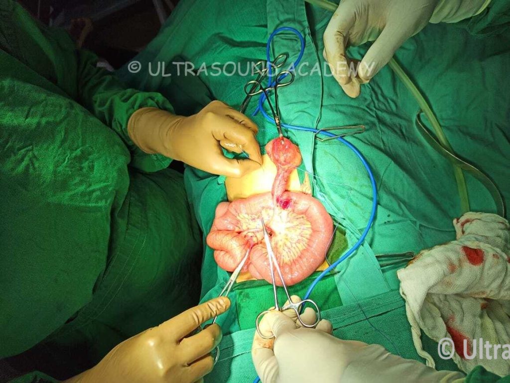
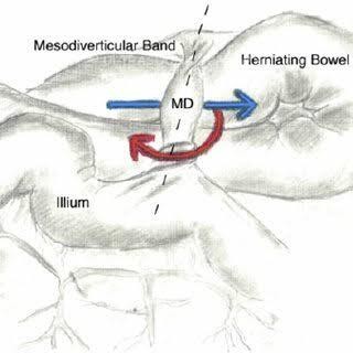
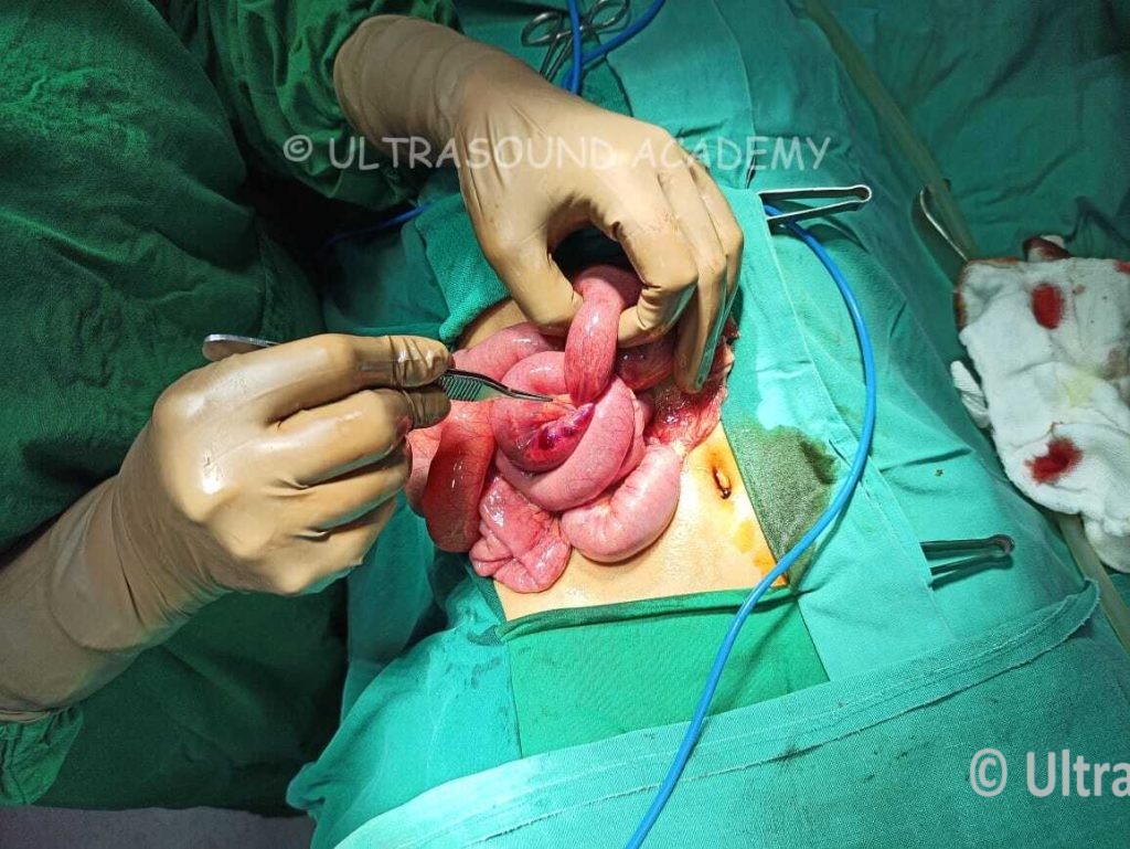
Meckel’s Diverticulitis with ileal volulus with mesodiverticular band confirmed on surgery.
Meckel’s diverticulitis can be challenging to diagnose on ultrasound (USG) due to its often nonspecific findings. However, in suspected cases, USG may reveal the following features:
Typical Ultrasound Findings:
- Blind-Ending Pouch:
A tubular or cystic structure located in the right lower abdomen, arising from the ileum.
It does not exhibit peristalsis like normal bowel loops.
- Wall Thickening:
Thickened walls of the diverticulum, suggesting inflammation.
May appear hypoechoic due to edema.
- Surrounding Inflammatory Changes:
Hyperechoic fat stranding around the diverticulum.
Possible localized fluid collection or abscess formation.
- Doppler Study:
Increased vascularity of the diverticular wall on color Doppler (hyperemia), indicative of inflammation.
- Intestinal Obstruction Signs:
Dilated bowel loops proximal to the diverticulum in cases where obstruction is associated.
- Free Fluid:
Presence of localized or free peritoneal fluid in the pelvis or around the diverticulum in cases of perforation.
Challenges with USG:
Small size and variable position of Meckel’s diverticulum can make it difficult to visualize.
It may be confused with appendicitis, Crohn’s disease, or other ileocecal conditions.
Additional Imaging:
If Meckel’s diverticulitis is suspected but not clearly visualized on USG, CT scan or Meckel’s scan (Technetium-99m pertechnetate) may be more sensitive for confirming the diagnosis.