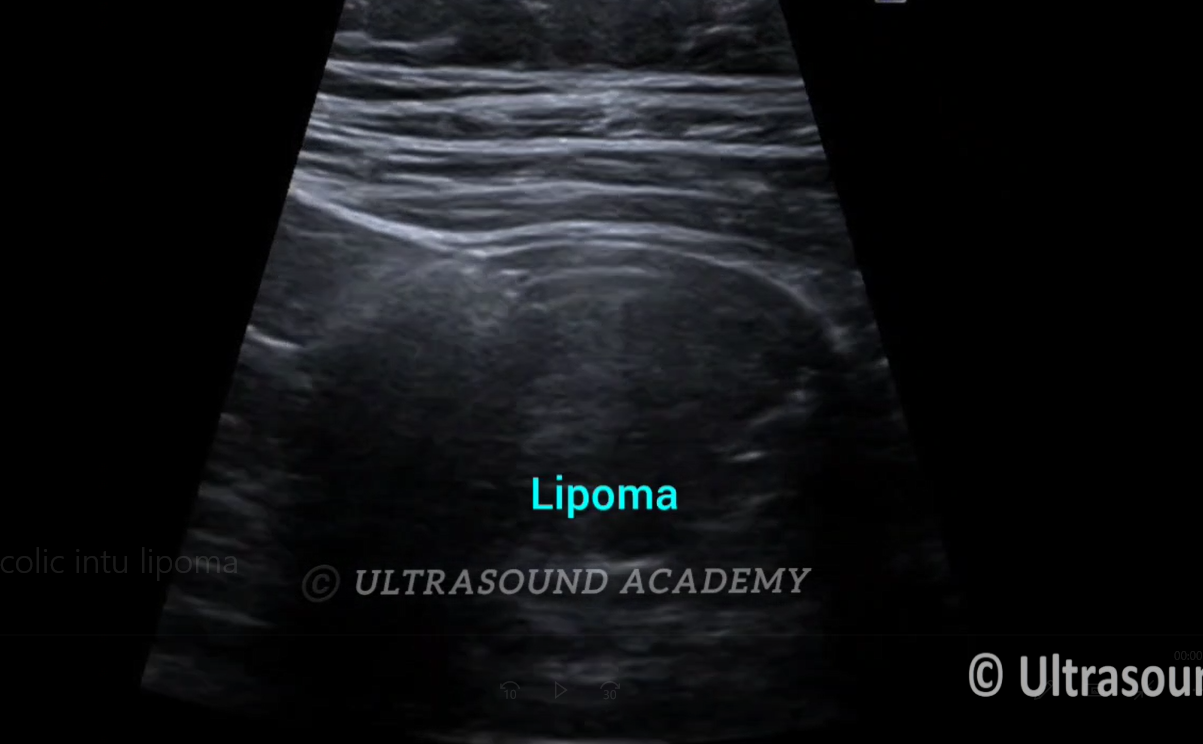
Findings and discussion- Elderly male having c/o left flank pain.
Telescoping of large bowel loop seen at left Iliac fossa S/O
Colo- Colic intussusception.
At lead point well defined oval hyperechoic lesion is seen.
This lesion appears to be lipoma.
No signs of bowel obstruction noted.
Bowel malignancy and lipoma are common caused of adult intussusception.
Adult intussusception is a rare condition where a part of the bowel telescopes into another bowel which can lead to bowel obstruction, ischemia, or perforation. Ultrasound is a useful imaging modality for diagnosing this condition, although it is more commonly used in children. In adults, the diagnosis is typically confirmed with a combination of clinical symptoms, radiologic findings, and sometimes, additional imaging such as CT scans.
On ultrasound, some common signs of intussusception include:
Target or Donut Sign: A classic finding, this appears as concentric rings or a “target” shape when the affected bowel segment is visualized in cross-section. This is due to the telescoping nature of the bowel layers.
Pseudo-kidney Sign: When viewed longitudinally, the intussuscepted bowel can resemble the appearance of a kidney, characterized by a thickened bowel wall and central hypoechoic areas.
Non-compressibility of Bowel Loops: The intussuscepted bowel may not compress when pressure is applied with the ultrasound probe, which can help differentiate it from other conditions.
Free Fluid: In cases of ischemia or bowel perforation, there may be free fluid or ascites around the affected area.
Absence of Peristalsis: The affected area may show decreased or absent bowel sounds when viewed with Doppler imaging, indicating a lack of normal bowel movement.
It is important to note that while ultrasound can provide initial evidence of intussusception, a CT scan is often required for more accurate assessment and confirmation of the diagnosis, especially in adults, due to its higher sensitivity.