Middle aged female having upper abdominal vague pain.
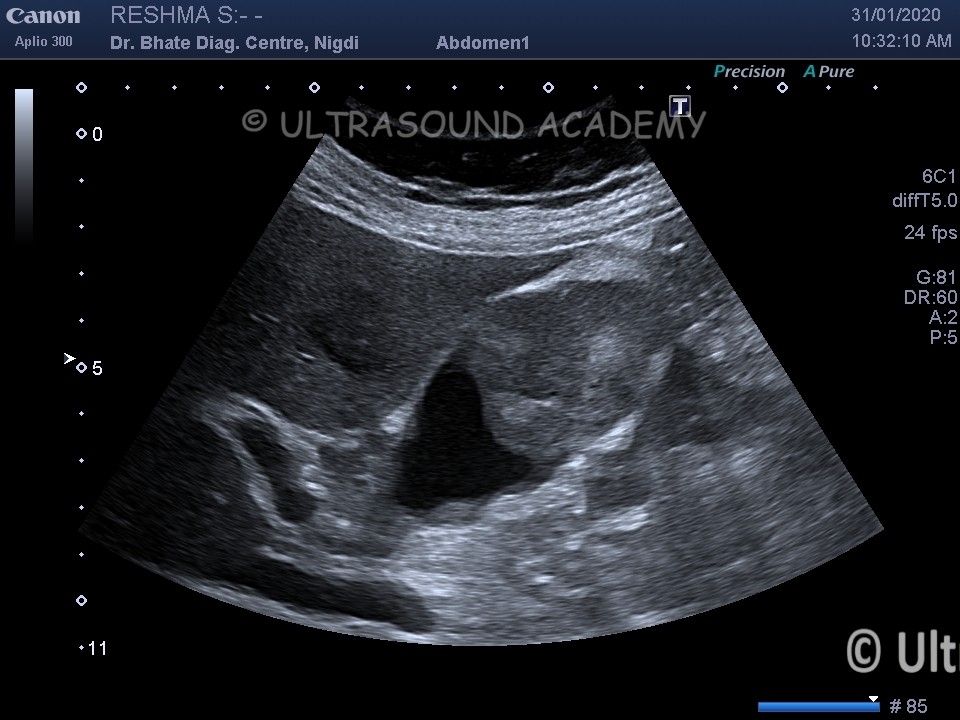
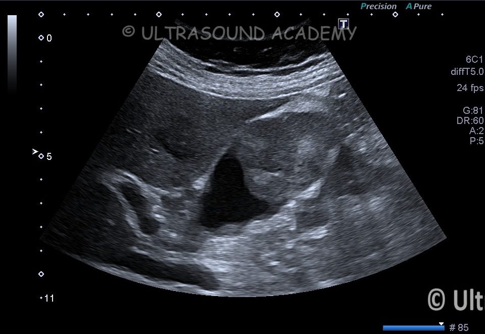
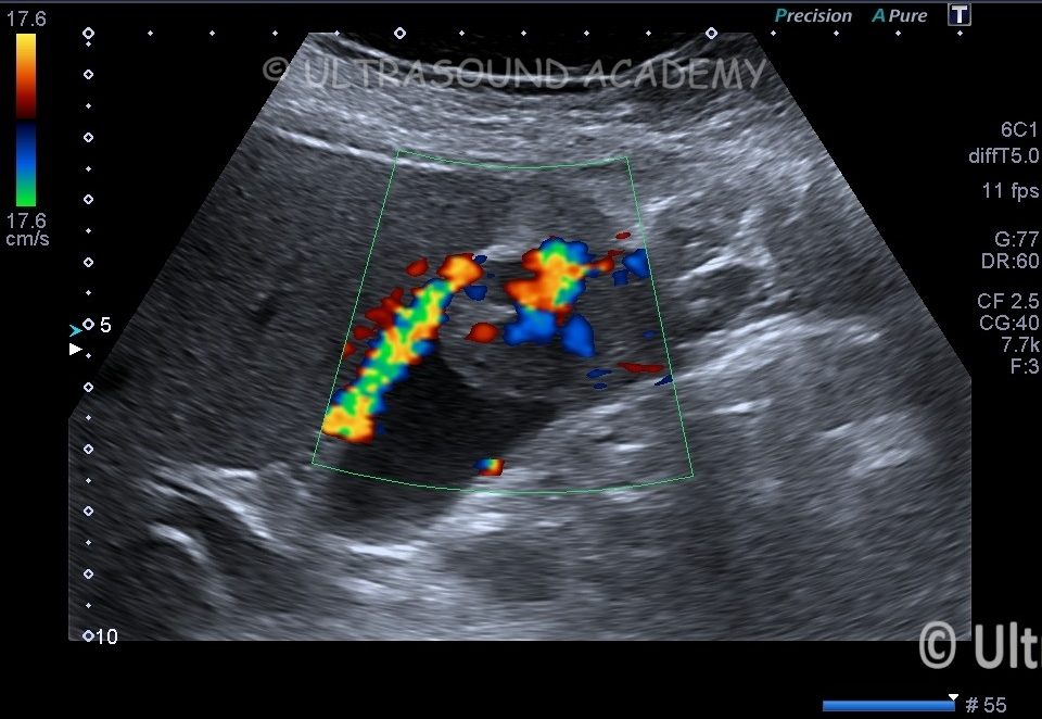
Large lobulated hypoechoic lesion is seen in GB near fundus.
significant vascularity is seen within the lesion.
No direct invasion of the adjacent liver parenchyma is noted.
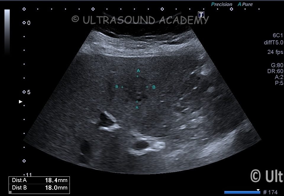
Well defined hypoechoic lesion is also seen in left lobe of liver. This lesion appears to be metastasis.
GB mass and liver mets confirmed on CECT.
On an ultrasound, carcinoma of the gallbladder (GBC) may present with various findings, depending on the stage of the disease. Common sonographic features include:
Focal or Diffuse Gallbladder Wall Thickening
Thickened wall with irregular margins.
Typically asymmetric in advanced cases.
Intraluminal Mass
A solid, irregular mass within the gallbladder lumen, often attached to the wall.
May show heterogeneous echotexture.
Loss of Normal Gallbladder Outline
The contour of the gallbladder may become disrupted due to tumor infiltration.
Gallstones
Present in 70-90% of cases, often associated with the carcinoma.
Direct Infiltration of Adjacent Organs
The liver, bile ducts, or other nearby structures may show evidence of invasion.
Pericholecystic Fluid
Fluid collection around the gallbladder due to inflammation or tumor spread.
Liver Involvement
Hypoechoic or hyperechoic lesions in the liver adjacent to the gallbladder suggest direct tumor extension or metastasis.
Lymphadenopathy
Enlarged lymph nodes in the porta hepatis, retroperitoneum, or other regions.
Dilated Biliary Tree
Dilatation of intrahepatic or extrahepatic bile ducts may occur if the tumor obstructs the bile flow.
A definitive diagnosis requires further imaging (CT/MRI) or biopsy to confirm malignancy.