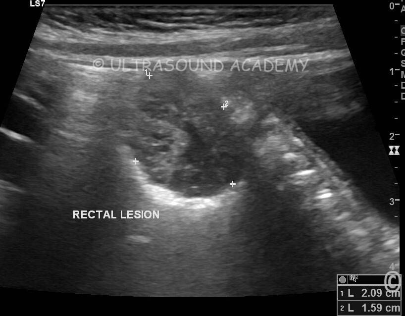
5 yr old child referred to USG with complaints of per rectal bleeding.
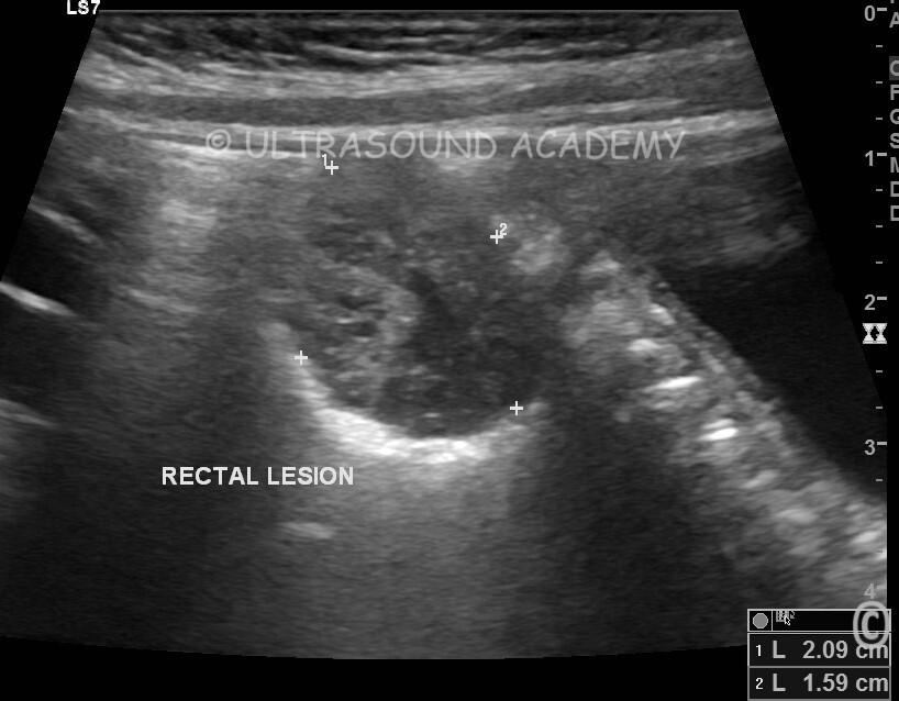
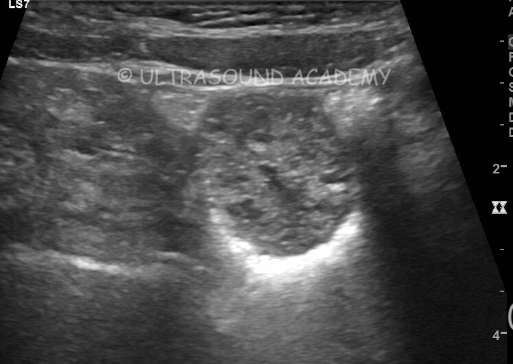
USG lower abdomen shows large hypoechoic lesion posterior to the urinary bladder.
This lesion seems to be within lumen of rectum.
Lesion shows few tiny cystic spaces.
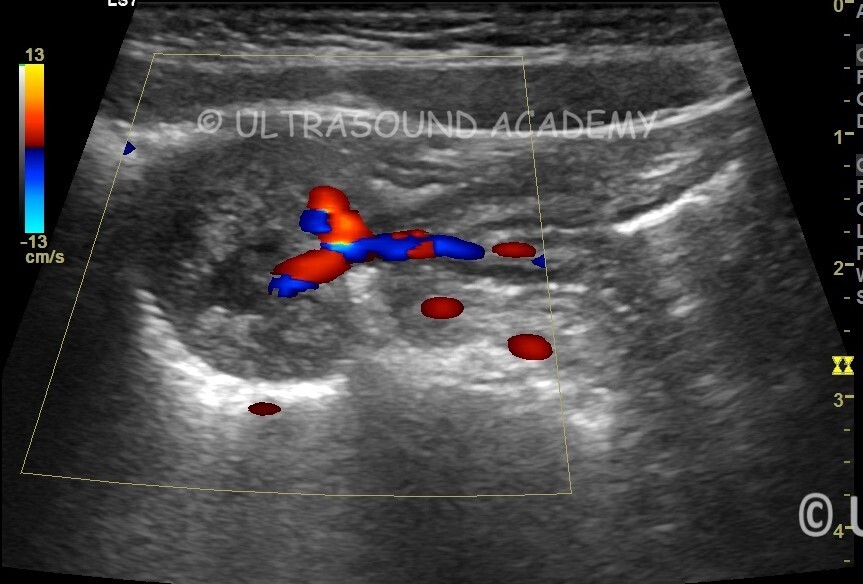
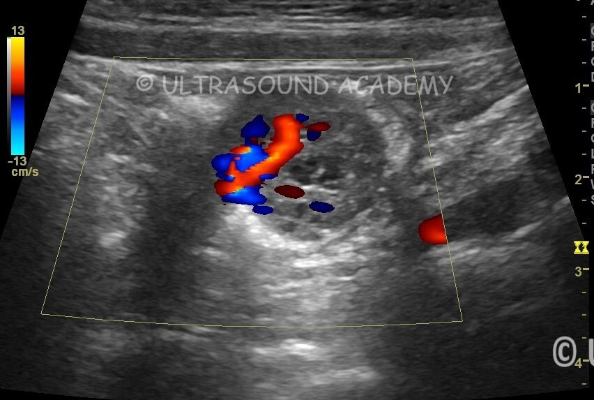
Sigficant vascularity is seen within the lesion.
Vascularity pattern is very typical and seen in radiating patten from the pedical of the lesion.
This lesion typical of rectal polyp.
Rectal Polyp in Children – Ultrasound (USG) Findings and Diagnosis
Rectal polyps in children are usually juvenile polyps, which are benign growths of the intestinal lining. They often present with painless rectal bleeding and may cause mucus in the stool.
Role of Ultrasound (USG):
While ultrasound is not the primary diagnostic tool, it can help assess complications such as intussusception or rectal wall thickening. More commonly, colonoscopy or sigmoidoscopy is used for direct visualization.
Ultrasound Findings:
Hypoechoic or Mixed-Echoic Mass: A well-defined, polypoid lesion may be seen in the rectal lumen.
Thickened Rectal Wall: If inflammation is present.
Doppler Study: Can help assess vascularity (polyp usually shows vascularity).
Intussusception Signs: If the polyp has caused bowel obstruction.