3-month-old female child with right inguinal swelling came for Ultrasound.
Findings and discussion:
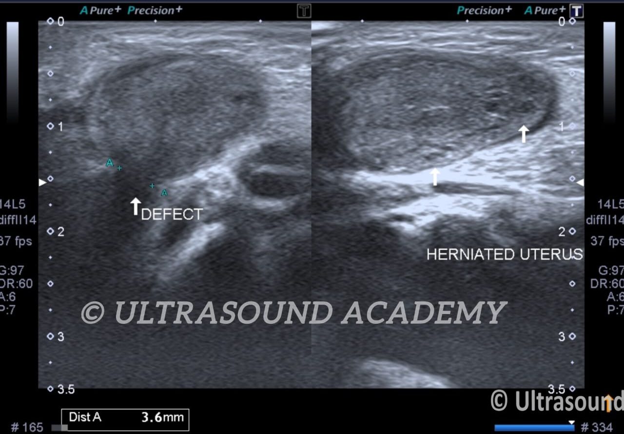
B mode USG shows herniation of uterus as well as left ovary into the inguinal canal. (Canal of Nuck hernia)
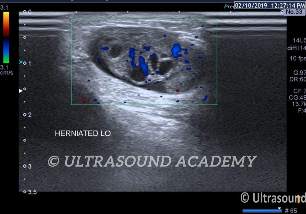
Uterus and ovary is normal in echotexture and show normal vascularity.
Right ovary is seen in the abdominal cavity.
Diagnosis of uterine inguinal hernia is made.
Key point: Ovarian inguinal hernia are prone to strangulation & ovarian torsion.
A uterine inguinal hernia is a rare condition, usually seen in female infants and young children. It involves herniation of the uterus, often along with one or both ovaries and fallopian tubes, into the inguinal canal. Ultrasound is the modality of choice for evaluating this condition. Here are the typical findings:
Ultrasound Findings
Identification of Uterus in the Inguinal Canal:
Hypoechoic or isoechoic structure with a central endometrial stripe (echogenic line), characteristic of the uterus.
The uterine shape (pear or tubular) is identified within the inguinal canal or hernia sac.
Ovarian Involvement:
The ovary may also be present in the inguinal canal.
Look for a round or oval structure with multiple small follicles distributed around its periphery (a classic ovarian appearance).
Doppler imaging should confirm blood flow to the ovary, ruling out torsion.
Fallopian Tubes:
Tubular structures may accompany the uterus and ovary in the hernia sac.
Hernia Sac and Inguinal Canal:
The hernia sac will be identified as an outpouching of peritoneum extending into the inguinal canal.
Dynamic ultrasound (e.g., with crying, Valsalva, or compression) may accentuate the hernia and aid diagnosis.
Associated Findings:
Ovarian torsion: Absence of blood flow to the ovary on Doppler.
Hydrocele or fluid: May be seen around the hernia sac or within the inguinal region.
Key Considerations:
Differential Diagnosis:
Differentiation from inguinal hernias containing bowel or other structures.
Differentiation from lymphadenopathy or other inguinal masses.
Confirmation of Blood Flow:
Always check for vascular compromise of the uterus and ovary using Doppler imaging to rule out ischemia.
Management Guidance:
Findings should be promptly reported to guide surgical consultation and intervention, especially if vascular compromise or torsion is suspected.
A uterine inguinal hernia is an uncommon condition, predominantly observed in female infants. It involves the protrusion of the uterus, and occasionally the ovaries and fallopian tubes, into the inguinal canal. Ultrasound imaging is the preferred diagnostic tool for evaluating this condition.
Ultrasound Findings:
Uterus in the Inguinal Canal:
Identification of the uterus within the inguinal canal is rare but has been documented. The uterus may appear as a mass in the inguinal region, and its presence can be confirmed through ultrasound imaging.
Ovarian Involvement:
The ovary may also be present in the inguinal canal. Ultrasound can detect the ovary within the hernia sac, and Doppler imaging should be used to assess blood flow, ensuring there is no vascular compromise.
Hernia Sac and Inguinal Canal:
Ultrasound can identify the hernia sac extending into the inguinal canal. Dynamic maneuvers, such as the Valsalva maneuver, can accentuate the hernia, aiding in diagnosis.
Key Considerations:
Differential Diagnosis:
Differentiation from other inguinal masses, such as lymphadenopathy or hydrocele of the canal of Nuck, is essential. Ultrasound imaging can assist in distinguishing these conditions.
Confirmation of Blood Flow:
Assessing vascular flow using Doppler imaging is crucial to rule out ovarian torsion or ischemia.
Management Guidance:
Prompt surgical consultation is recommended, especially if there is suspicion of vascular compromise or torsion, to prevent potential complications.