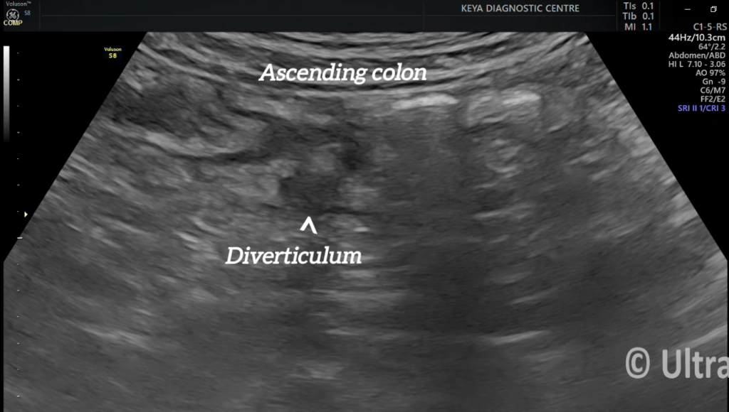
DIVERICULITIS – Case By Dr. Nitin Jadhav
45-year-old female with RIF pain, came for ultrasound.
Findings and discussion:
USG shows hypoechoic outpouching arising from the ascending colon.
The outpouching shows some degree of acoustic shadowing due to presence of gas or inspissated feces.
Bowel wall thickening is seen.
Surrounding echogenic inflamed fat is noted.
So, diagnosis of diverticulitis is made.
Diverticulitis can further complicate into:
Abscess, fistula formation, small bowel obstruction, perforation peritonitis or lower GI haemorrhage.
So, early and accurate diagnosis is important.
Ultrasound (USG) in Diverticulitis Diagnosis
Ultrasound (USG) is a useful, non-invasive imaging modality for diagnosing diverticulitis, especially when CT is unavailable or contraindicated (e.g., pregnancy, allergies to contrast). While CT remains the gold standard, ultrasound is increasingly recognized for its accuracy in detecting diverticulitis, particularly in experienced hands.
Key Ultrasound Findings in Diverticulitis
- Thickened Bowel Wall (>4 mm):
- Affected segment of the colon (commonly the sigmoid) appears thickened.
- Hypoechoic Inflamed Bowel Wall:
- Suggests edema and inflammation.
- Pericolic Fat Stranding:
- Appears as an echogenic area around the bowel due to inflammation.
- Diverticula:
- Small, round, hypoechoic outpouchings from the bowel wall, sometimes containing fecoliths.
- Abscess Formation:
- Seen as a hypoechoic fluid collection with possible gas bubbles.
- Hyperemia on Doppler Ultrasound:
- Increased blood flow around the inflamed segment suggests active inflammation.
- Free Fluid or Gas (Perforation Signs):
- Free air may be difficult to detect but can indicate perforation.
Advantages of Ultrasound in Diverticulitis
- Radiation-free (good for young patients and pregnant women).
- Portable and bedside-friendly.
- Can differentiate from other causes of abdominal pain (e.g., appendicitis, colitis).
Limitations
- Operator-dependent.
- Less accurate in obese patients.
- May not detect small abscesses or complications as well as CT.
Comparison with CT Scan
When is Ultrasound Recommended?
- First-line imaging in young patients, pregnant women, or where CT is unavailable.
- Follow-up imaging to monitor progress of known diverticulitis.
- Alternative to CT if contrast allergy or renal impairment is a concern.
If ultrasound suggests diverticulitis but is inconclusive, a CT scan is usually recommended for confirmation and staging.
error: Content is protected !!