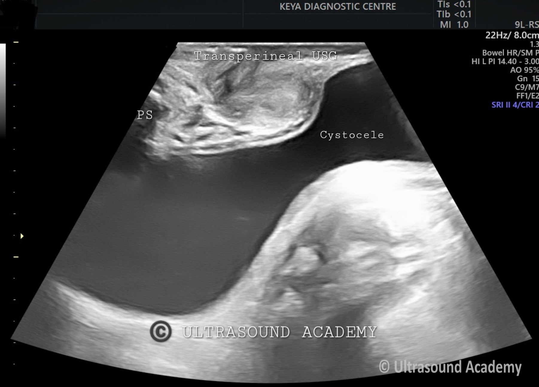
Elderly female complaining of mass coming out of vagina.
Findings :
Images and cine from transperineal ultrasound shows
Significant portion of urinary bladder descends down below the level of pubic symphysis on Valsalva s/o Cystocele.
Cystocele, or anterior vaginal wall prolapse, occurs when the bladder descends into the vaginal space due to weakened pelvic floor support. Transperineal ultrasound (TPUS) is a valuable, non-invasive imaging technique used to assess pelvic organ prolapse, including cystocele.
Prior pelvic surgery with suspected prolapse recurrence
Pre- and post-operative assessment of prolapse surgery
2. Ultrasound Technique
Patient Preparation
The patient should have a comfortably filled bladder (not overdistended).
The examination is performed in the dorsal lithotomy or standing position to assess prolapse dynamics.
Probe Placement
A low-frequency (3–6 MHz) convex or linear transducer is placed on the perineum, covered with gel and a protective sheath.
The transducer is oriented sagittally, allowing visualization of the pubic symphysis, bladder, urethra, vagina, rectum, and pelvic floor muscles.
3. Ultrasound Findings in Cystocele
Key Measurements & Criteria
Bladder Neck Descent (BND)
Measured as the movement of the bladder neck between rest and maximal strain.
BND > 10 mm is considered abnormal.
Retrovesical Angle (RVA)
The angle between the bladder base and the urethra.
RVA > 140° suggests significant prolapse.
Bladder Base Descent
A descent of the bladder base below the inferior margin of the pubic symphysis (commonly >10 mm below) is diagnostic of cystocele.
Hymenal Reference Line
The lowest point of the bladder descent is measured relative to the hymen.
If the bladder base descends below -10 mm, a cystocele is confirmed.
Urethral Hypermobility
Excessive rotation (>30°) or excessive movement of the urethra during straining.
4. Grading of Cystocele on TPUS
Mild (Stage 1): Bladder descent <10 mm below the pubic symphysis.
Moderate (Stage 2): Descent 10–20 mm below the pubic symphysis.
Severe (Stage 3-4): Descent >20 mm or prolapsing beyond the vaginal introitus.
5. Advantages of Transperineal Ultrasound
Non-invasive, well-tolerated
Real-time dynamic assessment (can assess prolapse during straining)
Superior to clinical examination for subtle prolapse
No radiation exposure
Useful for surgical planning (assesses pelvic support defects)
6. Limitations
Operator-dependent
May require MRI or urodynamic studies for a comprehensive evaluation in complex cases.
Conclusion
Transperineal ultrasound is an effective tool for assessing cystocele and other pelvic floor disorders. It provides objective, dynamic measurements that aid in diagnosis, severity assessment, and pre-surgical planning. It should be integrated with clinical examination and patient symptoms for optimal management.