Neonate with blackish skin discoloration Referred for ultrasound abdomen
Findings and discussion:
On B mode ultrasound:
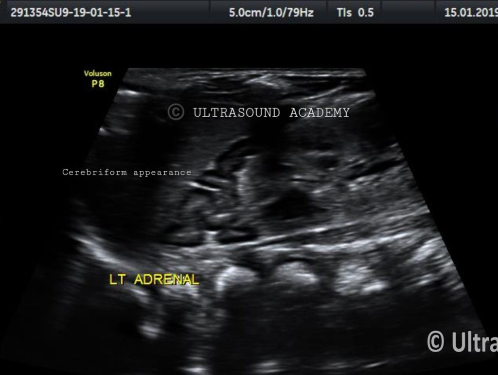
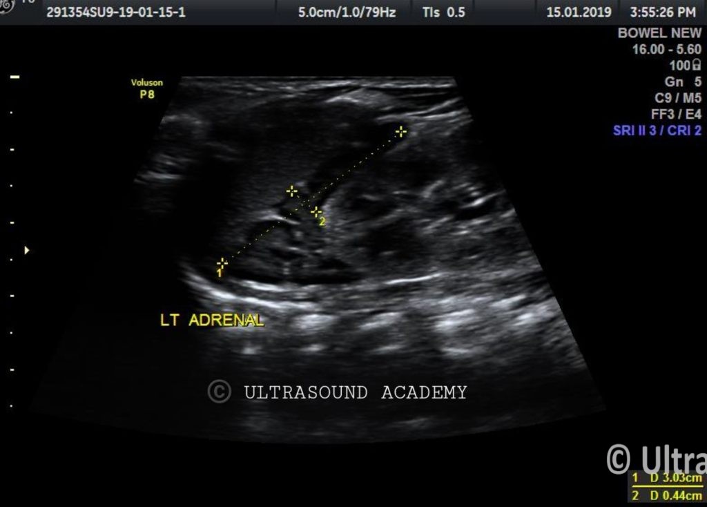
Left adrenal gland appears bulky and elongated and showed typical cerebriform pattern.
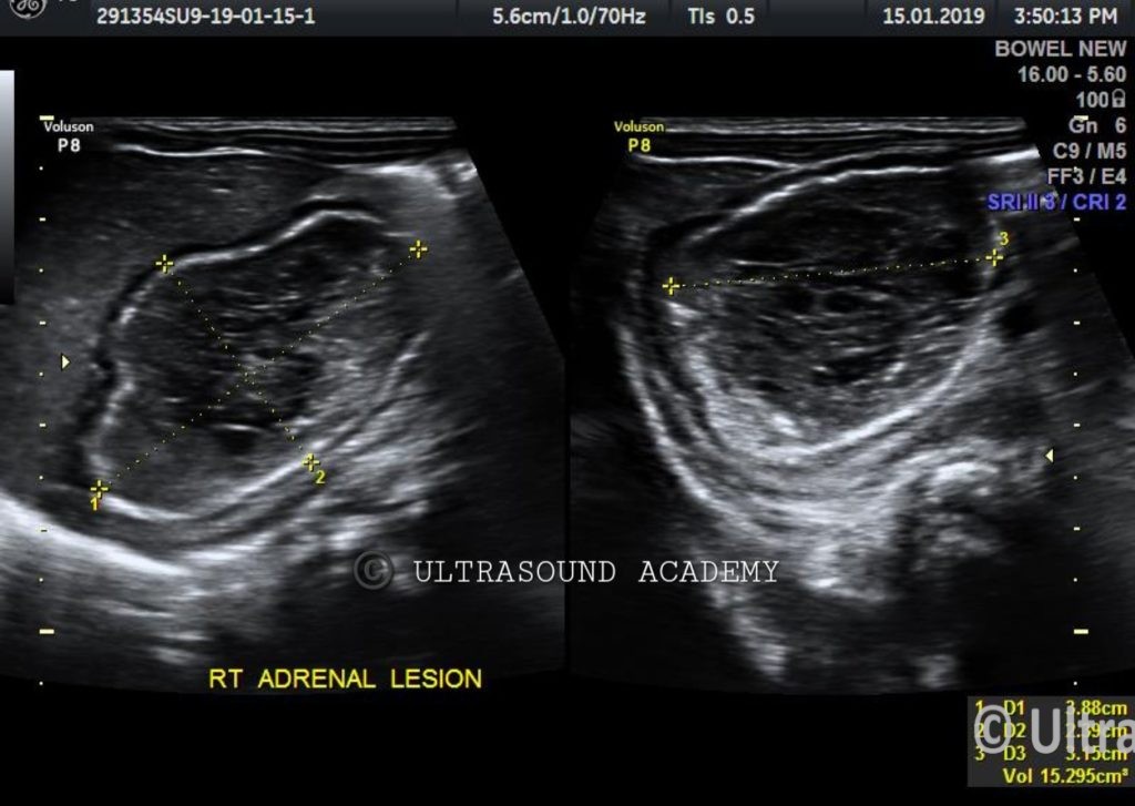
There is well defined heterogenous lesion is seen at right suprarenal region. This lesion showed calcified walls and internal hypoechoic septations representing debris/ blood products.
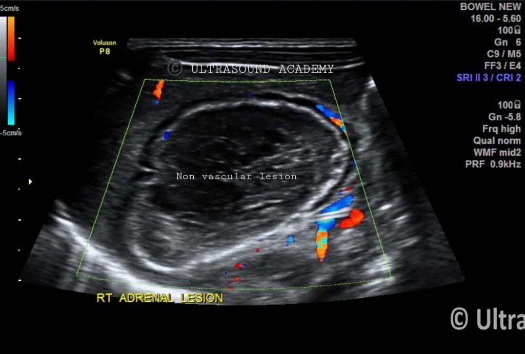
No significant vascularity or hyperaemia seen on Color Doppler study. Diagnosis of right adrenal haemorrhage is made.
The presence of calcification can be seen as early as 1-2 weeks after the onset.
So, the diagnosis of Left congenital adrenal hyperplasia and right adrenal haemorrhage is given.
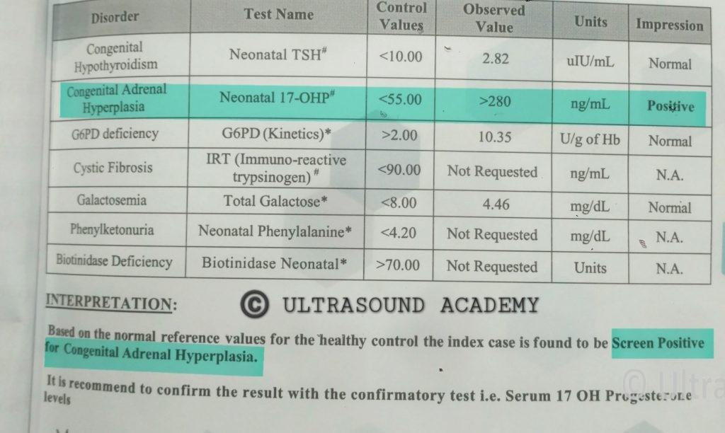
Neonatal screening test was done for CAH and it turned out to be positive for Neonatal 17 OH-Progesterone.
Congenital Adrenal Hyperplasia (CAH) and Ultrasonography (USG)
Ultrasound (USG) is often used as a diagnostic tool for Congenital Adrenal Hyperplasia (CAH), particularly in newborns and infants, to assess adrenal gland morphology and support clinical and laboratory findings.
Role of Ultrasound in CAH Diagnosis:
Adrenal Gland Enlargement
The adrenal glands appear bilaterally enlarged with a characteristic “cerebriform” or “lobulated” appearance due to hyperplasia of the adrenal cortex.
The increased size is a result of excess adrenocorticotropic hormone (ACTH) stimulation due to enzyme deficiencies (most commonly 21-hydroxylase deficiency).
Altered Adrenal Echotexture
The glands may show a hypoechoic or heterogeneous texture due to hyperplastic changes.
Adrenal medulla may remain relatively hyperechoic.
Sexual Differentiation Assessment (Ambiguous Genitalia)
In female infants with virilization, pelvic ultrasound can assess the presence of a uterus and ovaries to confirm the female reproductive anatomy.
In male infants with suspected salt-wasting CAH, USG is not primarily used, but can help rule out other differential diagnoses.
Screening for Associated Findings
Helps rule out adrenal tumors, adrenal hemorrhage, or cysts that may mimic CAH.
May detect signs of hydronephrosis or nephrocalcinosis due to excessive steroid treatment.
Fetal Ultrasonography (During Pregnancy)
In high-risk pregnancies, prenatal USG may detect adrenal hyperplasia and help in early intervention.
Combined with amniocentesis or chorionic villus sampling (CVS) for genetic testing to confirm the diagnosis.
Limitations of USG in CAH
Not a definitive diagnostic tool; it must be correlated with clinical symptoms, biochemical tests (17-hydroxyprogesterone, cortisol, electrolytes), and genetic testing.
USG findings may overlap with other adrenal disorders (tumors, hemorrhage, infections).
Other Imaging Modalities
MRI or CT scan may be used if detailed adrenal anatomy is needed, especially in atypical or late-diagnosed cases.