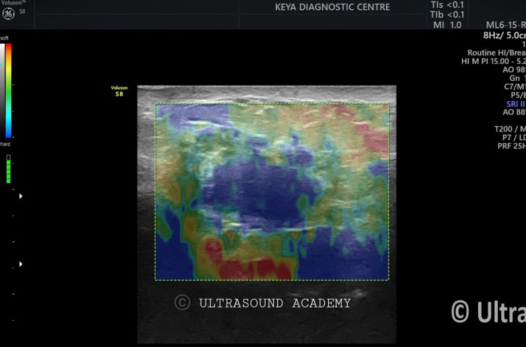
45 years old female referred by general surgeon for ultrasound breast in view of lump in right breast.
Findings and discussion:
On B mode ultrasound images and cine:
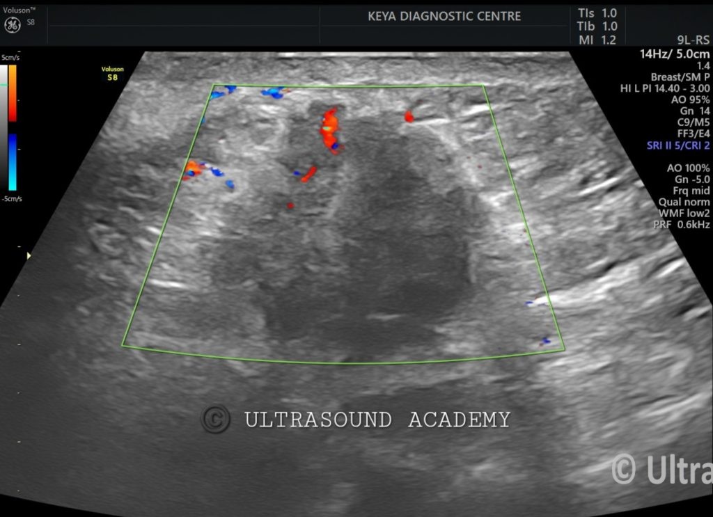
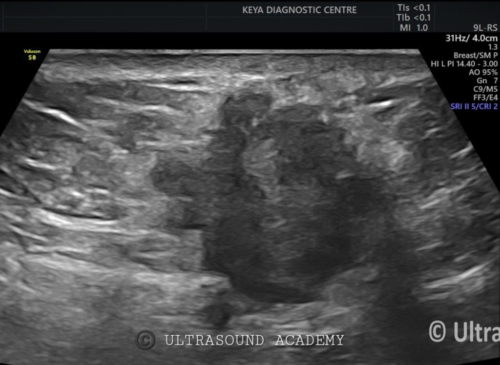
There is an ill-defined large lobulated heterogenous predominantly hypoechoic lesion is seen involving the upper and lower inner quadrant of right breast.
The lesion is taller than wider.
It showed some speculations.
Subtle specks of microcalcification seen.
Lesion shows surrounding echogenic halo suggestive of infiltration.
On Color Doppler it showed vascularity.
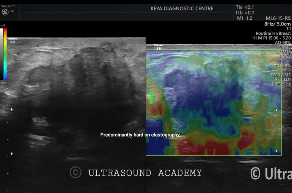
On elastography, the lesion is predominantly hard as shown by the blue Color.
On screening the abdomen:
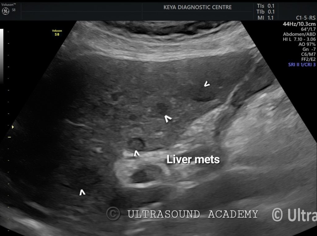
There are multiple well defined rounded heterogenous lesions of varying size, showing hypoechoic surrounding halo representing hepatic metastasis.
So, the diagnosis of breast mass with hepatic metastasis is given.
BI-RADS for Ultrasound (USG)
The Breast Imaging Reporting and Data System (BI-RADS) for ultrasound is a standardized classification system developed by the American College of Radiology (ACR) to assess breast lesions based on their ultrasound characteristics. This helps radiologists categorize findings, recommend appropriate follow-up, and guide clinical management.
Ultrasound Features in BI-RADS Classification
Benign Features (BI-RADS 2-3)
Well-defined, circumscribed margins
Oval or round shape
Hypoechoic, anechoic (cystic)
Posterior acoustic enhancement (common in cysts)
Parallel orientation
Suspicious Features (BI-RADS 4-5)
Irregular, spiculated, or microlobulated margins
Hypoechoic or heterogeneous echotexture
Taller-than-wide orientation
Posterior acoustic shadowing
Ductal extension or calcifications
Clinical Implications
BI-RADS 1 & 2: No concern, continue routine screening.
BI-RADS 3: Follow-up with repeat ultrasound in 6 months.
BI-RADS 4 & 5: Biopsy recommended.
BI-RADS 6: Malignancy confirmed, proceed with treatment.