BRAIN HEMORRAHAGE OR INFRACT– Case By Dr. Nitin Jadhav
Clinical history: 36 weeks of gestation patient came for routine growth scan. Findings:
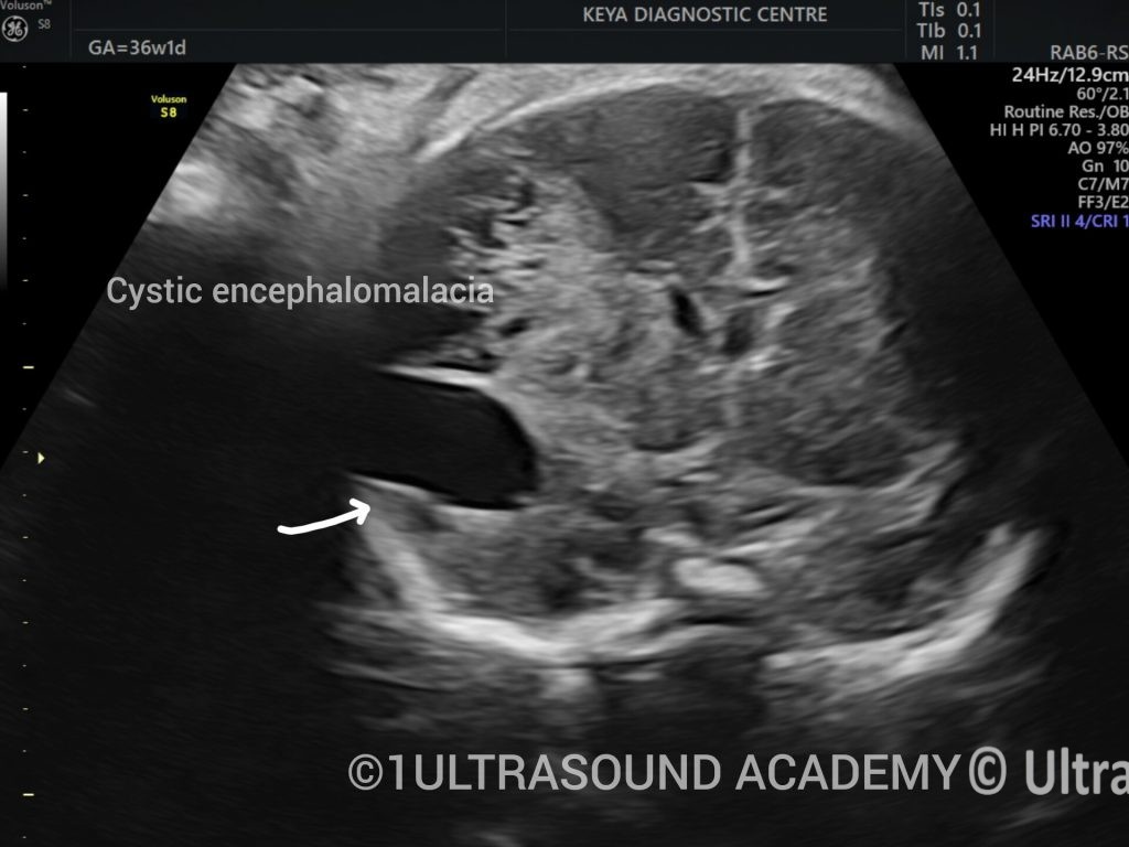
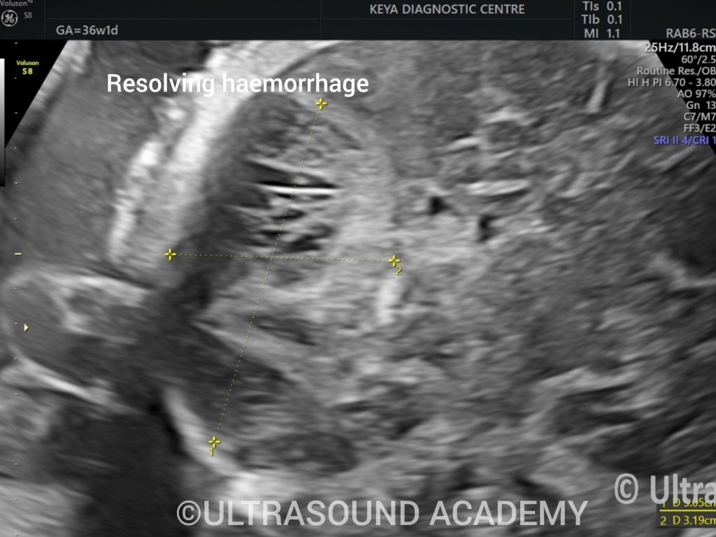
Approximately 5 x 3.1 cm sized large heterogeneous area is seen at left temporo-parietal region. This areas shows multiple tiny elongated cystic spaces within, largest cystic space measures 2.2 x 1.6 cm. No calcification is seen within the lesion. No vascularity is seen within the lesion. Approximately 0.9 x 0.4 cm sized small hyperechoic area is seen in the frontal horn of left ventricle.
All the above findings appears to be secondary to germinal matrix and brain parenchymal haemorrhage/infarct with intraventricular extension and leukomalacia.
Postnatal evaluation of brain is advisable.
Discussion:
Intracranial haemorrhage, vascular occlusion and infections or destructive lesions of the foetal brain.
Intracranial haemorrhage:
The periventricular germinal matrix in the foetus is highly vascular. The blood vessels are fragile with little stroma for support. In left 2nd and early 3rd trimester the vessels in the zone can bleed due to fluctuations in the foetal blood pressure resulting in germinal matrix haemorrhage. The germinal matrix haemorrhage may trigger terminal venous thrombosis resulting in venous infarct or intraparenchymal haemorrhage
Disposing factors are maternal, foetal and pregnancy related:
- Maternal Factors: Maternal allow immune thrombocytopenia, idiopathic thrombocytopenic purpura, von willebrand disease, thrombophilia, systemic lupus erythematous, Vitamin K deficiency, trauma, drugs like warfarin and antiepileptics
- Foetal coagulopathies, anaemia, inborn error of metabolism, gene mutations associated with familial intraparenchymal haemorrhage and infections
- Perinatal hypoxia and asphyxia, placental abruption, preeclampsia, chorioamnionitis and complications of monochorionic twinning such as twin to twin transfusion and co twin demise are of obstetric complications that may cause foetal intracranial haemorrhage.
Grading of ICH
Grade I- confined to germinal matrix haemorrhage. Normal lateral ventricles
Grade II- intraventricular bleed filling < 50% of lateral ventricle, normal lateral ventricle size
Grade III- intraventricular bleed feeling more than 50% of the lateral ventricle, ventriculomegaly
Grade IV- grade I, II or 3 with parenchymal bleed
GMH is located in the periventricular germinal matrix add the caudothalamic groove. During the 1st 3-8 days (acute phase), the haemorrhage is hyperechoic. In the following 2 weeks (subacute phase), the centre of the haemorrhage becomes hypoechoic surrounded by hyperechoic rim. Eventually (chronic phase), it reacts, is sharply demarcated and uniformly hypoechoic. This evaluation applies to haemorrhage in any location
Intraventricular haemorrhage is suspected is any of the following findings are seen:
- Bulky an irregular shaped choroid plexus
- Hyperechoic content in the lateral ventricle- the choroid plexus, anatomically it does not extend anterior to the foramen of monro, hence a hyperechoic lesion in the anterior horn is clot
- Irregular, thickened and hyperechoic ventricular margins
- Particulate CSF in the ventricle with dependant debris
E intraventricular adhesions or synechiae
- Lateral ventriculomegaly due to obstruction of the foramina of monro
E regression of ventricular size with resolution of haemorrhage
error: Content is protected !!