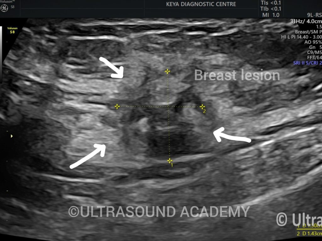
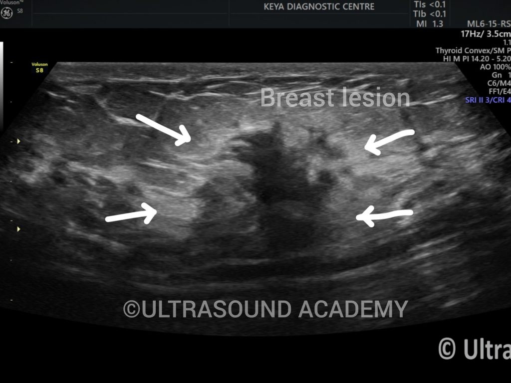
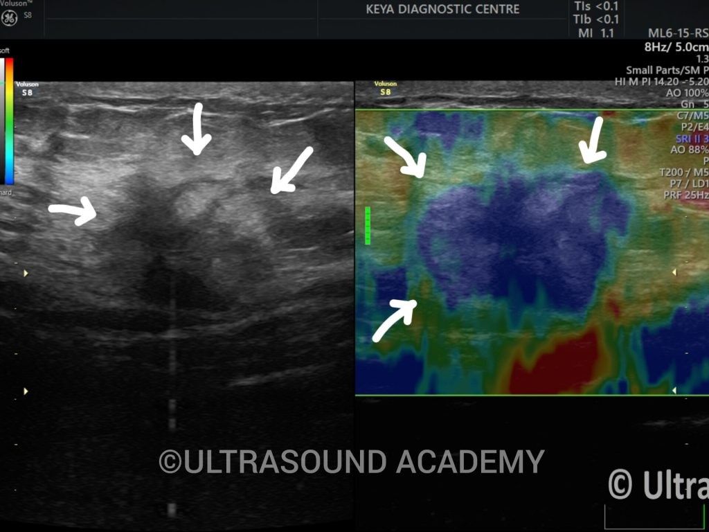
An elderly female presented with complaints of painless, hard, lump in the breast.
Findings and Discussion:
On 2D ultrasonography, an ill-defined oval shaped heterogeneously hypoechoic lesion with irregular lobulated margins, non-parallel orientation. On colour doppler lesion shows increased vascularity.
However, no obvious intralesional calcification was evident on ultrasound.
On strain elastography lesion shows blue hue on strain elastography. It suggest lesion is hard inconsistency.
These findings were in favour of malignant breast lesion and diagnosis of BI-RADS V ( highly suggestive of malignancy)
A BI-RADS (Breast Imaging Reporting and Data System) 5 lesion indicates a high suspicion of malignancy. This category is used in breast imaging reports, particularly from mammograms, ultrasounds, and MRIs, and it suggests that the lesion has characteristics strongly indicative of breast cancer.
Key points regarding BI-RADS 5 lesions:
Implication: A BI-RADS 5 classification means that there is a greater than 95% likelihood that the lesion is cancerous.
Follow-Up: Immediate further evaluation is typically recommended, which may include a biopsy to confirm the diagnosis
Lesion Characteristics: BI-RADS 5 lesions might have certain features, such as irregular shapes, spiculated margins, and microcalcifications, that help radiologists determine the likelihood of breast cancer.
Management: If a biopsy confirms malignancy, the findings will influence treatment decisions, which may include surgery, radiation, chemotherapy, or hormone therapy, depending on the specifics of the diagnosi
If you or someone you know has received a BI-RADS 5 diagnosis, it’s important to discuss the results and next steps with a healthcare provider for appropriate management and care.
Key Points:
BI-RADS provided lexicon for both ultrasound and mammography to characterise and categorised a breast lesion from benign to malignant. BI-RADS lexicon can be found in further readings.