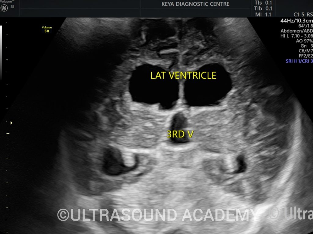
Clinical History: Patient referred for routine growth scan
Findings
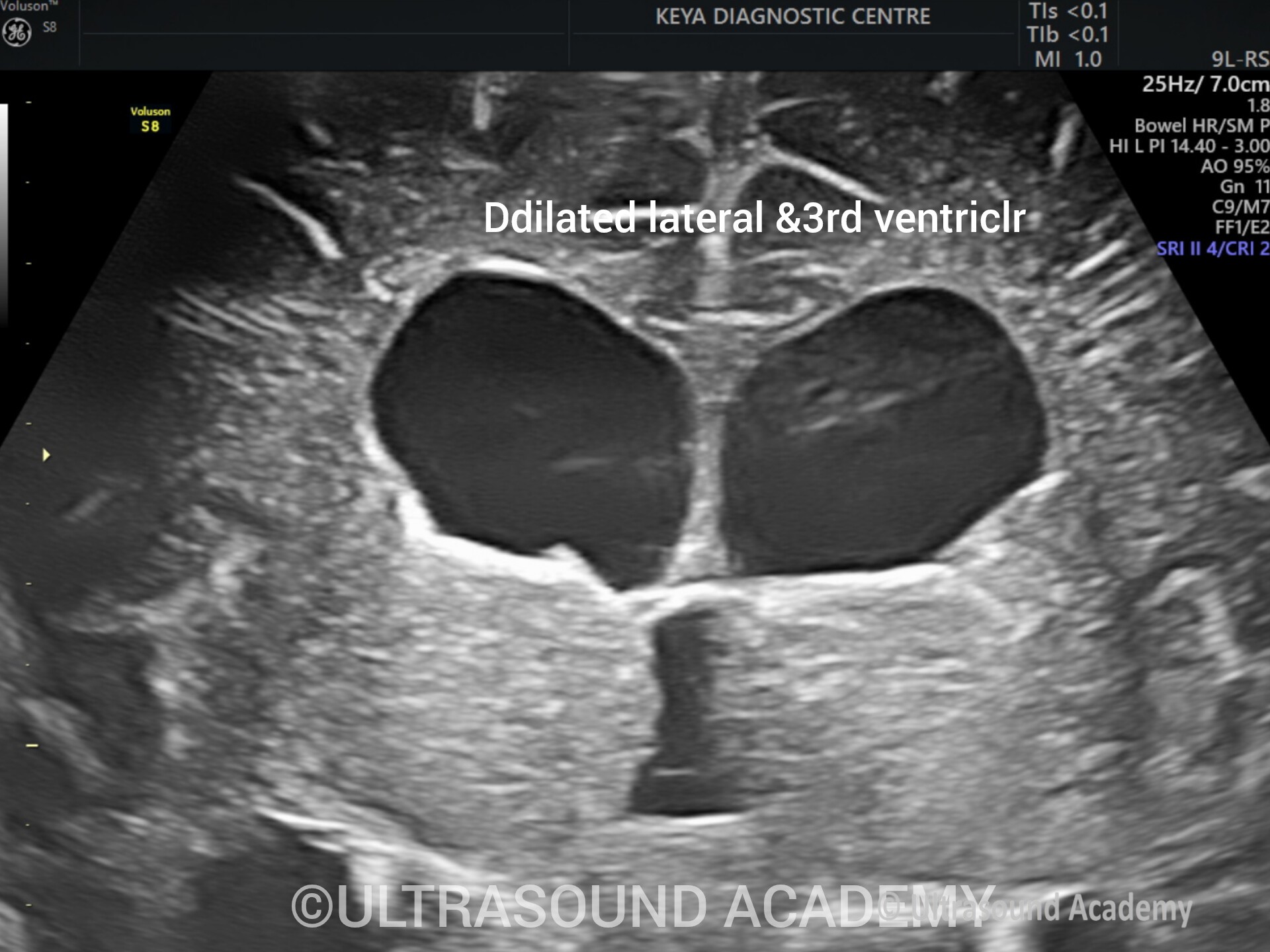
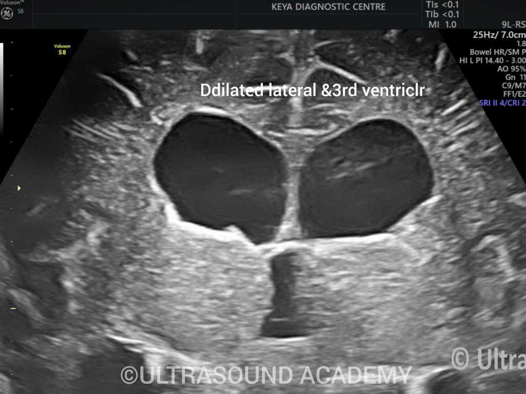
Both lateral ventricles of brain are significantly dilated.
Third ventricle also appears dilated and fourth ventricle appears and dilated
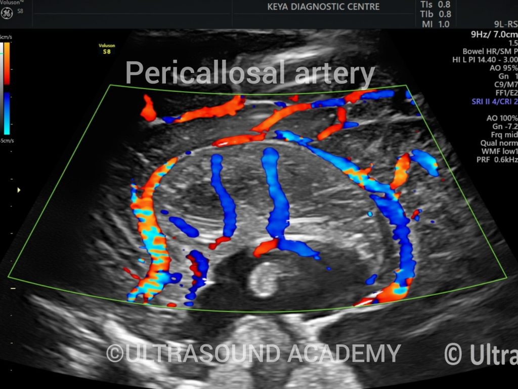
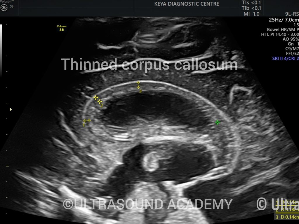
Corpus callosum is well visualised
Posterior fossa brain appears normal
No other obvious foetal anomaly is noted.
Possibility of aqueductal stenosis is likely. Postnatal detailed evaluation with MRI is advisable
Discussion
Antenatal aqueductal stenosis refers to a narrowing of the aqueduct of Sylvius, a narrow channel that connects the third and fourth ventricles of the brain. This condition, which can be detected during prenatal ultrasounds, leads to impaired cerebrospinal fluid (CSF) flow between these ventricles and may result in hydrocephalus (accumulation of CSF in the brain).
Key Points About Antenatal Aqueductal Stenosis:
Definition:
Aqueductal stenosis is a congenital narrowing of the aqueduct of Sylvius. This blockage or narrowing prevents normal CSF circulation and can lead to the dilation of the ventricles above the blockage (known as ventriculomegaly) and hydrocephalus.
It is the most common cause of non-communicating hydrocephalus in fetuses.
Causes:
Aqueductal stenosis may be genetic or acquired. Most cases are idiopathic (unknown cause), but several factors can contribute to the condition:
Genetic factors: In some cases, aqueductal stenosis is inherited in an autosomal recessive or X-linked pattern.
Prenatal infections: Infections during pregnancy (such as toxoplasmosis, cytomegalovirus (CMV), or rubella) may lead to aqueductal stenosis.
Structural brain malformations: It can be associated with other structural abnormalities of the brain, including Chiari malformation, Dandy-Walker malformation, and Aicardi syndrome.
Congenital malformations: Some syndromes, such as neuronal migration disorders or holoprosencephaly, may be associated with aqueductal stenosis.
Diagnosis:
Aqueductal stenosis is typically detected antenatally through imaging during a routine prenatal ultrasound:
Prenatal ultrasound: If aqueductal stenosis causes hydrocephalus or ventriculomegaly, it may be detected in the second trimester. The ultrasound shows dilation of the ventricles (usually the lateral ventricles) and sometimes an enlarged third ventricle.
Fetal MRI: If the prenatal ultrasound suggests ventriculomegaly or hydrocephalus, a fetal MRI may be used to get a more detailed image of the brain’s structure and confirm the presence of aqueductal stenosis and any associated abnormalities.
Postnatal imaging: After birth, cranial ultrasound or MRI can be used to assess the degree of ventriculomegaly and any potential structural brain defects that may be associated with aqueductal stenosis.
Signs and Symptoms:
Prenatal signs: The primary sign of aqueductal stenosis during pregnancy is the dilation of the fetal ventricles. This may be accompanied by oligohydramnios (low amniotic fluid) in some cases.
Postnatal signs: After birth, if the condition leads to hydrocephalus, the infant may exhibit signs such as:
Enlarged head circumference
Bulging fontanel (soft spot on the head)
Severe developmental delay
Seizures
Poor feeding and irritability
Motor impairments or hypotonia (reduced muscle tone)
Complications:
Hydrocephalus: The primary complication of aqueductal stenosis is the accumulation of cerebrospinal fluid, leading to hydrocephalus. If left untreated, hydrocephalus can cause brain damage and developmental delays.
Brain damage: Chronic pressure from accumulating CSF can damage brain tissues, leading to cognitive impairment, motor deficits, and other neurological issues.
Neurological deficits: The severity of the condition and its impact on the baby’s development depend on the degree of obstruction and the timing of intervention.